What’s actually slowing this PC down?
Pick the symptom - the matching free tool is one click away.
Some links on this page are affiliate links: if you buy through them we may earn a commission, at no extra cost to you.
Neither CRISPR activation (CRISPRa) nor transcription-factor overexpression is a proven universal winner for turning skin fibroblasts into neurons. CRISPRa uses a DNA-binding but non-cutting dCas9 activator plus guide RNAs to switch on the cell’s own neuronal genes. Classical direct conversion supplies neuronal regulators such as ASCL1, BRN2 (POU3F2) and MYT1L from external expression constructs. Both approaches can engage the same gene-regulatory circuits, but they differ in how those circuits are reached.
A 2026 mouse study introduced mechanically enhanced CRISPR activation (MECA): brief mechanical deformation was used to make chromatin more accessible before CRISPRa targeted endogenous genes. The study reported approximately 3% neuronal conversion with CRISPRa alone, 10% with mechanical priming plus CRISPRa, and 40% after an OCT4-associated priming step followed by neuronal activation. Those are results from one mouse-fibroblast study, not a head-to-head proof that CRISPRa beats transcription factors or a demonstration in human cells. Human fibroblast-to-neuron conversion has been reported with transcription factors, including a study that reached about 16% under a combined factor, small-molecule and microRNA protocol.
As an Amazon Associate I earn from qualifying purchases.
Do these 3 things before closing this tab:
1Repair Windows errors before they cause bigger problems2Scan for outdated or missing drivers - takes under a minute3Clear out junk files and repair common Windows errorsWhat exactly is being compared?
CRISPRa and transcription-factor (TF) overexpression are two ways to trigger direct conversion, in which a differentiated fibroblast is pushed toward a neuronal identity without first passing through a pluripotent stem-cell state. The distinction is what the experiment delivers and where the regulatory proteins act.
CRISPRa activates endogenous genes
CRISPRa uses a catalytically inactive Cas9 protein (dCas9) fused to transcriptional activation domains. Guide RNAs direct that complex to regulatory DNA near a chosen gene, where the activator recruits the cell’s own transcription machinery. In the MECA study, modified RNA was used to deliver a dCas9-based synergistic activation mediator (SAM), and truncated guide RNAs were selected to avoid creating DNA double-strand breaks. Targets included endogenous Ascl1 and Brn2, so the fibroblast’s native gene copies, rather than replacement coding sequences, were activated.
#1 Best Overall

- PVC Material: Our human neuron anatomy model is made of imported non-toxic PVC material and is finely crafted. Each important part is clearly distinguished by color to ensure that each component is accurately reproduced.
- Digital Markings: Each part of the neuron anatomy model is clearly labeled with numbers, showing the anatomical structures of nerve cell axons, dendrites, Nissl bodies, neurofilaments, etc., and is accompanied by explanation cards that perfectly describe important information about neuron anatomy.
- Best Teaching Tool: This neuron anatomy magnification model is convenient to store and easy to clean. It can be used not only as a learning and teaching tool for medical students, but also as an intuitive teaching tool for learning human neuron cell anatomy in classroom laboratories. It is the best teaching tool for learning.
- Wide Application: This 2500x magnified neuron cell anatomy model is a highly realistic teaching model suitable for human anatomy research. Ideal for a doctor's office, classroom, or as a learning aid, this model provides a more inclusive educational experience.
- Clear Structure: Our neuron anatomy model, magnified 2500 times, clearly shows the anatomical details of the neuron cell structure. It is compact, durable, and easy to store. This anatomy model is designed to assist in dissection and teaching.
That “endogenous” feature does not make CRISPRa biologically unrelated to TF conversion. If a guide turns on the cell’s own ASCL1 gene, the resulting ASCL1 protein is still a lineage regulator. CRISPRa changes how the regulator is produced and controlled; it does not eliminate the need for neuronal transcription factors.
TF overexpression supplies the regulators
In classical direct conversion, researchers introduce expression constructs for neuronal lineage factors. The best-known mouse combination is BAM: BRN2, ASCL1 and MYT1L. These proteins can activate neuronal programs and, in the case of pioneering factors, help open otherwise inaccessible chromatin. Human fibroblasts often need a different cocktail, dose, timing or additional cues because conversion and maturation are more difficult in adult human cells.
The Tool Desk
Outbyte PC Repair FREEClear out junk files and repair common Windows errorsFree Scan →Outbyte Driver Updater FREEScan for outdated or missing drivers - takes under a minuteDriver Scan →Both strategies can bypass pluripotency, which may shorten the route to induced neurons and avoid some risks associated with an intermediate stem-cell state. Bypassing that state does not guarantee mature, subtype-accurate neurons; identity, function and yield still depend on the protocol.
How does MECA combine CRISPRa with mechanical priming?
MECA adds a physical chromatin-opening step to programmable gene activation. Song and colleagues first mapped how brief deformation changed chromatin accessibility in primary adult mouse fibroblasts, then used those maps to choose guide-RNA target regions. The workflow can be understood as four linked stages:
- Prime the cells mechanically. A short deformation treatment changes the physical state of the nucleus and temporarily exposes regulatory regions that are less accessible in untreated fibroblasts.
- Map the accessible regulatory landscape. Multi-omics measurements identify regions that become more open after the mechanical stimulus. These data are used to avoid selecting guide sites solely from a static DNA sequence.
- Activate neuronal fate genes. Modified RNA delivers the dCas9-SAM activator, while truncated guide RNAs bring it to endogenous targets such as Ascl1 and Brn2. Because dCas9 is non-cutting, the design is intended to activate transcription without the double-strand breaks made by nuclease-active Cas9.
- Test stronger priming combinations. The UCLA account also describes an OCT4-based priming step followed by neuronal-gene activation. In that mouse-cell context, the combination produced a substantially higher reported conversion fraction than mechanical priming with CRISPRa alone.
The central idea is that a guide cannot activate a regulatory site efficiently if the site is physically inaccessible. Song Li, the study’s senior author, described this as a “last-mile” accessibility problem: reprogramming factors may be present, yet unable to reach the DNA elements they must bind.
How does transcription-factor overexpression convert fibroblasts?
TF conversion starts by choosing a factor set that supplies the regulatory instructions for a desired neuronal subtype. The BAM combination—BRN2, ASCL1 and MYT1L—has converted mouse fibroblasts into induced neurons. In human cells, factor requirements are species- and subtype-dependent; a human fibroblast study used BRN2, MYT1L and FEZF2 to produce excitatory cortical neuron-like cells.
Rank #2
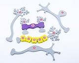
- Distinguish between multipolar, bipolar, unipolar neurons and interneurons
- Add myelin sheath pieces to explore two types of neuroglia in our nervous systems
- Examine the overall effect on neuronal firing at excitatory and inhibitory synapses
Why factor cocktails are not interchangeable
ASCL1 can initiate neuronal gene programs, BRN2 helps specify neuronal identity, and MYT1L suppresses competing non-neuronal programs. FEZF2 is associated with corticofugal and deep-layer cortical identities, so replacing one factor with another can change the neuronal subtype rather than merely changing efficiency. Reviews emphasize that a cocktail that works in mouse embryonic or young fibroblasts may perform differently in adult human donor cells.
What the human cortical-neuron study showed
The 2017 human-fibroblast study reported functional excitatory cortical neuron-like cells using BRN2, MYT1L and FEZF2. Adding small molecules and microRNAs raised the reported conversion efficiency to approximately 16% in that study’s protocol. The cells integrated into human neural networks, but that percentage is not a universal benchmark and cannot be compared directly with the MECA figures because the species, starting cells, factor combination, delivery system, subtype and assay differ. Read the primary human-fibroblast study.
What conversion efficiencies have actually been reported?
The published numbers come from different experimental designs. The table keeps the conditions attached to each percentage so they are not mistaken for a single competition.
| Approach and condition | Reported conversion | What the number represents |
|---|---|---|
| CRISPRa alone | About 3% | Mouse fibroblasts in the 2026 MECA study, before mechanical priming. |
| Mechanical priming plus CRISPRa | About 10% | Mouse fibroblasts receiving the brief deformation treatment and dCas9-based activation. |
| OCT4-associated priming followed by neuronal activation | About 40% | Mouse fibroblasts in the same study’s stronger priming sequence; not a human result. |
| BRN2, MYT1L and FEZF2 with small molecules and microRNAs | About 16% | Human fibroblast-to-excitatory-cortical-neuron protocol reported in 2017. |
The MECA values are summarized by UCLA’s October 22, 2026 account of the study, while the 16% value comes from the authors of the human cortical-neuron study. They should not be ranked as if all four conditions were tested side by side. A “converted cell” may also be defined differently between experiments, for example by marker expression, morphology, neuronal function or a combination of tests.
For the original MECA report, see Song et al. in Matter (2026) and the UCLA study summary published October 22, 2026.
Does CRISPRa outperform transcription-factor overexpression?
No matched evidence in the cited studies establishes a general winner. CRISPRa’s approximately 10% or 40% MECA-associated values cannot be used to claim superiority over TF conversion because they were obtained in mouse fibroblasts under specific priming and assay conditions, whereas the approximately 16% TF result was obtained in human fibroblasts directed toward excitatory cortical neurons.
Rank #3
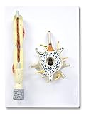
- 2,500X Enlarged Model of a typical Neuron
- An excellent model to study the internal structures of the cell body
- The myelin sheath with nodes of Ranvier and Schwann cells can be revealed by removing a part of the dendrite
- Mounted on a base
- Perfect for students and teachers
| Decision factor | CRISPRa / MECA | TF overexpression |
|---|---|---|
| What is delivered | dCas9 activator plus guide RNAs; MECA adds transient mechanical priming. | External genes or expression constructs encoding neuronal regulators. |
| Where activation occurs | At selected endogenous loci, with guide placement chosen around accessible regulatory regions. | Supplied factor proteins act on many downstream genes and regulatory elements. |
| Programming style | Guide sequences can be redesigned to address different endogenous genes. | Changing the factor cocktail, expression level or timing changes the fate program. |
| DNA cutting | The MECA design uses dCas9 and truncated guides intended to avoid double-strand breaks. | Whether DNA is cut depends on the delivery system; the cited TF studies are expression-based rather than nuclease-based. |
| Evidence highlighted here | New mouse-fibroblast MECA study; human performance remains untested in the cited account. | Multiple direct-conversion studies, including a human cortical-neuron protocol. |
| Main uncertainty | Whether accessibility-guided activation reaches comparable efficiency, maturation and subtype fidelity in human adult fibroblasts. | Whether a selected cocktail gives the intended subtype and mature function across donors and species. |
CRISPRa is therefore best viewed as a programmable way to turn on lineage regulators, not as a replacement for transcription-factor biology. TF overexpression remains a direct route with a larger body of conversion work, while CRISPRa offers finer control over which endogenous loci are activated and when.
Why is chromatin accessibility the key issue?
Fibroblast chromatin is organized for a connective-tissue program, not a neuronal one. A transcriptional activator can be present but ineffective if the enhancer or promoter it needs is buried in relatively closed chromatin. Mechanical deformation appears to alter that physical landscape temporarily, and the MECA study used multi-omics data to identify the regions most responsive to the treatment.
This is also why conventional TF cocktails can work without directly editing every neuronal gene: pioneering factors may help open chromatin and recruit additional regulators. CRISPRa and TF overexpression can thus converge on the same barrier from opposite directions—CRISPRa selects and activates endogenous loci, while supplied factors can both bind regulatory DNA and propagate a broader network response.
What are the main practical trade-offs?
Programmability versus simplicity
CRISPRa requires a dCas9 activator, guide-RNA design and a delivery schedule. Adding MECA introduces a mechanical-priming step and an accessibility-mapping workflow. A TF protocol may be conceptually simpler once a validated cocktail is known, but changing neuronal subtype usually requires testing a different combination of factors.
Endogenous regulation versus external expression
Activating the native ASCL1 or BRN2 locus preserves the cell’s own gene architecture and regulatory context. External constructs can produce strong, controllable expression, yet their level and duration may not match endogenous timing. Neither design automatically guarantees correct maturation or complete suppression of fibroblast programs.
Delivery and dose
The MECA report used modified RNA delivery for the dCas9-SAM system and truncated guides. TF studies use expression constructs and may add small molecules or microRNAs. Delivery efficiency, expression duration and cell toxicity can influence the apparent conversion percentage as much as the choice of biological regulators.
Windows Errors? Fix Them Before They Spread
Repair common Windows errors and clear accumulated junk for a smoother, more stable PC - no reinstall needed.Free scan · no reinstallOutdated Drivers Are Slowing You Down
One free scan finds every outdated or missing driver and matches the right update for your exact hardware.Free scan · exact hardware matchRank #4

- Professional Anatomical Teaching Aid: This life-size human brain model is designed specifically for medical professionals, educators, and college-level students. It serves as a precise visual aid for advanced neurological instruction, clinical patient communication, and surgical planning discussions
- Superior PVC Construction: Crafted from high-grade, corrosion-resistant PVC, this durable brain model ensures long-term reliability in rigorous professional environments. Its non-toxic, sturdy build is engineered for frequent handling by adults in laboratory and university settings, resisting oxidation for years of use
- 1:1 Scale with Base: Accurately scaled to real human brain proportions, this model measures 8*6.7*5.9 inches (20.5*17*15 CM) and includes a sturdy white base for stable display, allowing for clear identification of all major anatomical structures during professional demonstrations
- Removable 2-Part Design: Featuring a detachable two-part configuration, this model allows for easy separation to reveal internal structures. This design is ideal for instructor-led anatomical presentations and detailed study, facilitating a deeper understanding of the brain's complex regions without the need for dissection
- Comprehensive Functional Anatomy: This model distinctly maps critical regions including the frontal, parietal, occipital, and temporal lobes. It is an indispensable professional resource for medical institutions and universities seeking to illustrate the functional divisions of the human brain accurately
Subtype and maturation
A high marker-positive fraction is not the same as a mature, subtype-faithful neuron. Human adult-cell conversion can be less efficient, and induced-neuron identity can remain uncertain when it is inferred from a limited marker panel. Functional tests and, where relevant, integration into neural networks are needed to support claims beyond morphology.
Independent reader supportYour contribution helps us test, update, and keep practical guides available for everyone.How should a fair CRISPRa-versus-TF experiment be designed?
A meaningful comparison keeps the biological and measurement conditions aligned. At minimum, report the following variables for both methods:
- Species and donor age: mouse, fetal human or adult human fibroblasts can have very different epigenetic barriers.
- Fibroblast source: tissue origin, passage number and culture state affect baseline plasticity.
- Neuronal subtype: a generic induced neuron, an excitatory cortical neuron and another subtype are not interchangeable endpoints.
- Factor or guide set: list every activated gene, supplied TF, small molecule and microRNA, including timing and dose.
- Delivery method: distinguish modified RNA, viral or nonviral expression systems and the duration of expression.
- Priming treatment: specify deformation conditions or any OCT4-associated step rather than grouping all “CRISPRa” experiments together.
- Conversion definition: state whether the percentage is based on neuronal markers, morphology, survival, electrophysiological properties or network integration.
- Follow-up time: early marker expression and later maturation measure different outcomes.
Without these details, a larger percentage may simply reflect a more permissive donor, an easier subtype, a longer maturation period or a broader definition of conversion.
What is known about human-cell use and clinical readiness?
Human fibroblast conversion with TFs is established at the proof-of-concept research level, including the cortical-neuron study described above. That does not mean every adult donor converts efficiently or that the resulting cells are interchangeable with native neurons. Reviews identify species-dependent factor requirements, difficult maturation and unresolved questions about subtype fidelity.
Recommended Free Tools
MECA remains preclinical mouse research in the cited material. UCLA reports that the team plans to test conversion in human cells and determine whether the resulting neurons function adequately for applications such as drug testing or transplantation. Those are future investigations, not evidence that MECA currently works in human tissue or is ready for transplantation. No cited source establishes clinical efficacy, safety or human therapeutic benefit.
The broader field’s caution is summarized in the review “Evaluating cell reprogramming, differentiation and conversion technologies in neuroscience” and the PubMed-indexed review “Reprogramming of somatic cells: iPS and iN cells”. A later review also discusses the limits of inferring neuronal identity from markers alone: “The iNs and Outs of Direct Reprogramming to Induced Neurons”.
Best Value

- It displays the structure of a typical neuron.
- This item, magnified 2,500 times.
- The sectioned soma shows all the internal cellular organs including nucleus, Golgi complex, smooth and rough endoplasmic reticulum.
- The dendrite can be opened into two parts to reveal the internal structures such as the Schwann cell and myelin sheath.
- Environmentally friendly PVC, imported paint, computer color matching, hand-painted.
Which approach makes sense for a particular research goal?
Choose based on the biological question rather than the headline percentage.
- Use CRISPRa when endogenous regulation is central. It is suited to testing whether activating specific native loci, singly or in combinations, can initiate a fate program. MECA is especially relevant when chromatin accessibility is suspected to be the limiting step.
- Use TF overexpression when a validated lineage cocktail is the starting point. BAM and related combinations provide a direct way to test induced-neuron generation, while human cortical-neuron work shows that additional small molecules and microRNAs can materially change the outcome.
- Use matched parallel arms when efficiency is the question. Keep species, donor, subtype, delivery, timing and conversion criteria identical, then compare yield, maturation and function separately.
- Use hybrid designs when the biology supports them. CRISPRa can activate endogenous TF genes, while supplied factors or physical priming can address accessibility. Such combinations should be treated as new protocols requiring their own controls, not as proof that one platform alone is superior.
The most defensible conclusion is that CRISPRa and TF overexpression are complementary tools. MECA makes CRISPRa more responsive to chromatin state in mouse fibroblasts, but human performance and functional suitability remain open questions; TF conversion has human evidence but still depends strongly on donor, subtype and maturation conditions.
Frequently Asked Questions
Does CRISPRa permanently edit the fibroblast’s DNA?
The MECA system uses catalytically inactive dCas9 rather than a nuclease, and its truncated guides are designed to avoid double-strand breaks. That means the reported mechanism is transcriptional activation, not sequence cutting, although guide-dependent and delivery-related effects still need to be evaluated experimentally.
Can CRISPRa activate a neuron subtype without supplying any transcription factors?
It can target endogenous transcription-factor genes, so the cell may produce its own ASCL1, BRN2 or other regulators after guide delivery. In biological terms, those factors still drive the conversion; CRISPRa changes how they are induced rather than making them unnecessary.
Why might a mouse protocol fail in adult human fibroblasts?
Species, donor age and chromatin state alter which regulatory sites are accessible and which factor combinations are sufficient. Human adult cells may also mature more slowly or require additional subtype-specific cues, so mouse conversion percentages cannot be transferred directly.
What proves that an induced cell is really a functional neuron?
A convincing assessment combines neuronal identity with functional behavior, rather than relying on one marker. The human cortical-neuron study went beyond marker expression by reporting excitatory neuron-like cells that integrated into human neural networks, an endpoint that a simple morphology count cannot establish.
Quick Recap
Product prices and availability are accurate as of the date/time indicated and are subject to change. Any price and availability information displayed on Amazon at the time of purchase will apply.

