Do these 3 things before closing this tab:
1Fix the driver behind crashes, sound loss and screen glitches2Repair Windows errors before they cause bigger problems3Scan for outdated or missing drivers - takes under a minuteA 2008 live-cell imaging study used luminescent platinum(II) complexes to make cellular structures easier to distinguish from background. Because the probes kept emitting for microseconds after excitation, researchers could delay image capture until much of the faster cellular autofluorescence had faded. The work demonstrated a laboratory imaging method—not a clinical tool or consumer lighting product.
What the 2008 study demonstrated
The paper, “Time-resolved and two-photon emission imaging microscopy of live cells with inert platinum complexes,” explored time-resolved emission imaging microscopy (TREM) in living cells. Its probes were small, charge-neutral platinum(II) complexes in the [PtLCl] family. A platinum(II) complex is a molecule built around a platinum ion in the +2 oxidation state; here, the complexes were designed to emit light after excitation and to accumulate inside cells.


As an Amazon Associate I earn from qualifying purchases.
The authors reported intracellular accumulation after a five-minute incubation and demonstrated imaging in three cell types: normal human dermal fibroblasts, neoplastic C8161 cells, and CHO cells. They also reported preferential localization to nucleic-acid structures, particularly nucleoli. These findings describe the tested cells and experimental conditions, not a general targeting guarantee.
Free tools Windows power users keep installed
One-click scans. No signup required.
The primary paper reported emission quantum yields of up to 70%. Quantum yield describes how efficiently absorbed energy is re-emitted as light. The authors also described the complexes as having low cytotoxicity under the studied conditions; that observation does not establish broad safety, and it is not evidence of safety in people.
#1 Best Overall
Why a longer emission lifetime helps
Waiting for background light to fade
Cellular autofluorescence is light emitted by molecules naturally present in cells after excitation. In the paper, conventional fluorophores and relevant background signals are described as having lifetimes of a few nanoseconds, while the platinum complexes emit on microsecond timescales. A luminescence lifetime is the time an excited molecule continues emitting after it has been stimulated.
TREM uses that difference in timing. The instrument excites the sample, waits briefly, and then opens a detection window. By the time the camera records the image, much of the short-lived background has decayed, while the longer-lived platinum signal remains. This can improve contrast by rejecting some background light; it does not mean every source of image noise disappears.
Rank #2
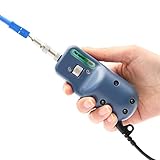
- [●400 times magnification, the fiber end face is clearly visible]
- [●Less than 1μm resolution, clear imaging.]
- [●USB Port compatible with the PCs and Laptops(Windows), OTDR( Komshine)]
- [●Applicable for UPC: SC/FC/ ST/LC/ SC Female/LC Female(Included); Applicable for APC: SC/SC Female(Optional) ]
- [●Quick focus manually. Light weight and portable, simple operation.]
How it differs from ordinary fluorescence imaging
| Aspect | Conventional fluorescence described in the paper | Platinum-complex TREM demonstration |
|---|---|---|
| Emission lifetime | A few nanoseconds | Microsecond-scale emission |
| Background handling | Short-lived signal and cellular autofluorescence can overlap in time | Delayed detection can suppress short-lived background after it fades |
| Cell demonstrations | Not specified as a particular comparison set in the cited paper | Normal human dermal fibroblasts, neoplastic C8161 cells, and CHO cells |
Chemistry World’s 2008 coverage characterized the probe as glowing hundreds of times longer than conventional probes. Its accessible explanation contrasts several microseconds with a few nanoseconds. Treat that as a description of this reported work, not as a universal performance specification for imaging probes.
Two-photon excitation extended the demonstration
The study also used two-photon excitation (TPE), in which a molecule absorbs two lower-energy photons nearly simultaneously to reach an excited state. The authors combined the approach with near-infrared two-photon excitation. This is a distinct excitation mode from the usual one-photon approach; the paper presents it as part of the laboratory demonstration rather than proof of a clinical imaging capability.
Rank #3
What the findings do—and do not—establish
- Established in the paper: microsecond-scale emission, quantum yields up to 70%, intracellular accumulation after a five-minute incubation, and imaging in the three reported cell types.
- Limited to the reported conditions: the characterization of low cytotoxicity and the observed intracellular localization. These results do not amount to validated safety across doses, tissues, or people.
- Proposed, not demonstrated as an established application: deeper tissue imaging and antibody conjugation. The paper discussed antibody conjugation as a possible extension. Chemistry World reported at the time that the probe had not yet been coupled to biological molecules.
- Not established by these sources: current commercial availability, clinical adoption, human use, or compatibility with a particular microscope or accessory.
The study is a 2008 laboratory research demonstration. Its authors showed how emission lifetime can provide another source of contrast in live-cell microscopy, but the cited evidence does not establish a currently marketed product or a clinical method.
Quick Recap
Best Value

- 1. 400 times Fiber Optic Connector Inspection Probe and 3.5 inch HD LCD display.
- 2. Fiber Optic Connector Inspection Probe Suit for FC/SC/LC/ST UPC Male Connect Head and SC/LC UPC Female Connect Head
- 3. Small size, one-handed, convenient and fast.Rotary metal bracket, stable and convenient.
- 4. 4000mAH large capacity lithium power, 20 hours standby.
- 5. Real time video,support extend t-flash card,quick check the pollution degr
Sources
- Botchway et al., “Time-resolved and two-photon emission imaging microscopy of live cells with inert platinum complexes,” Proceedings of the National Academy of Sciences, 2008.
- Chemistry World, October 15, 2008, report on platinum-based dyes for cell imaging.
- Chemistry World, October 29, 2008, explanation of the probes’ longer emission and the then-unrealized biological conjugation.
Product prices and availability are accurate as of the date/time indicated and are subject to change. Any price and availability information displayed on Amazon at the time of purchase will apply.




