Recommended Free Tools
A 2025 study used defocused spatially-offset Raman spectroscopy (SORS) to observe how experimental drug depots formed and released drugs beneath porcine skin. The laser measured molecular signals; it did not trigger release. The work demonstrates a research method in an ex vivo model, not a proven way to monitor implants in patients.
How the laser technique works
Raman spectroscopy detects molecular vibrations from which researchers can identify chemical signals. In spatially-offset Raman spectroscopy, the light-collection point is displaced from the point where the laser illuminates the sample. That offset helps gather information from below the surface. Rath and colleagues used a defocused SORS arrangement to monitor an in situ forming implant (ISFI) beneath skin without labeling the model drugs. The study, published in the Journal of Controlled Release in 2025, describes the measurements as non-invasive in its experimental skin model.
As an Amazon Associate I earn from qualifying purchases.
What the researchers tested
The team placed full-thickness porcine skin in a custom flow-through diffusion cell and studied two model drugs with different solubility characteristics: hydrophilic 4-cyanophenol (4-CP) and hydrophobic all-trans retinoic acid (RA). SORS monitored the subcutaneous implant, while high-performance liquid chromatography (HPLC) quantified drug that entered the receptor medium. Confocal Raman microscopy images of cross-sections were used to check the SORS findings, and static Franz diffusion-cell experiments served as a reference for the flow-through measurements.
Crashes, No Sound, or Screen Glitches?
Random freezes, missing sound and display glitches usually trace back to one bad driver. Find and replace yours safely.Free scan · under a minutePC Slower Than It Used to Be?
A free scan shows the junk files, broken settings and background clutter dragging Windows down - then fixes them in one click.Free scan · Windows 10 & 11What happened to the two model drugs
| Model drug | Released after 2.5 days | What the result means |
|---|---|---|
| Hydrophilic 4-cyanophenol (4-CP) | 90.7% in static conditions; 94.8% in flow-through conditions | Pronounced early release in the study’s model formulations and experimental setup. |
| Hydrophobic all-trans retinoic acid (RA) | 3.3% in static conditions; 2.1% in flow-through conditions | Release was much lower over the same period under the reported conditions. |
These are results for the study’s formulations, not typical release rates for implants generally and not patient outcomes. The authors linked implant formation with release behavior and identified solvent exchange as a key driver of the burst observed for 4-CP. The two compounds’ contrasting results show why monitoring an implant’s changing composition and measuring drug released into surrounding fluid can provide complementary information.
#1 Best Overall
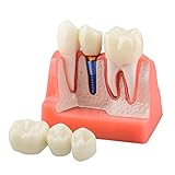
- Crowns are made of high-quality resin material. The implants and abutments are made of metal and not cheap plastic. High quality materials ensure that the display model is sturdy and sturdy.
- Crowns, roots, implants and abutments can be separated, which makes it easier to demonstrate the tooth structure individually to patients. Magnets embedded in the crown and the top of the root ensure ease of detachment. The magnets also make the crown and the root attach firmly in place with virtually no risk of falling off.
- The 4 times dental implant model comes with a separate bridge to better educate patients about implant and bridge restorative options. The 4X Model visually helps patients better understand dental restoration procedures.
- The cross-section base of the gingival bone is convenient for doctors to show the tooth structure to the patient. It also shows the integral relationship between the implant and the root.
- The dental implant model is an excellent tool to introduce and explain dental implant(s) to patients or students. The excellent visual explanation makes it easier for everyone to understand.
What the results establish—and what they do not
The study shows that SORS can be used alongside chemical quantification to characterize experimental implant formation and model-drug release in an ex vivo porcine-skin setup. It supports further work on formulation development and the possibility of individualized therapeutic drug monitoring, but it does not establish that the technique works in people, improves clinical decisions, has regulatory clearance, or is commercially available. The reported evidence is from an experimental model, not a clinical monitoring trial.
Observing release is not triggering it
SORS was used to observe molecular signals associated with implant formation and drug release; the study does not show that its laser controls or accelerates release. A separate 2021 study investigated a purpose-designed ocular implant in which pulsed near-infrared light triggered release from a PLGA capsule containing light-activated liposomes. That distinct approach was studied in vitro and in vivo, and should not be conflated with the 2025 SORS work. Read the separate ocular-implant study.
Rank #2

- Educational Model: Dental Typodont implant transparent model with removable mandibular base implant, which can show the implant more intuitively.
- Real Materials: The dental implant material is acrylic resin, the transparent base, and the implant screws are also solid materials, which can show the mandibular overdenture more realistically.
- Educational Tool: The dental implant restoration model is an excellent educational tool for dental students, dentists, and implant training education.
- Durable Structure: The entire model is made of real dental implant material, which makes the model durable and reusable, and an excellent choice for demonstration.
- Multiple Applications: It is suitable for dentists to show visual effects more intuitively in communication, and is used to demonstrate dental implant placement techniques. It is a perfect gift for dentists.
How this approach differs from other imaging research
Other studies have examined implant release using different signals and sample setups; they are not direct, head-to-head comparisons with SORS.
- A 2022 MALDI mass-spectrometry imaging paper mapped active pharmaceutical ingredient distribution in non-conductive long-acting polymer implants and examined release.
- A 2012 MALDI-TOF imaging study examined controlled-release lipid implants, reporting drug-rich domains and concentration gradients.
- A 2020 UV-visible imaging study examined early leuprolide release and implant formation in laboratory matrices intended to emulate the subcutaneous environment.
These methods answer related but different questions. To compare them, consider what chemical signal each detects, whether measurements are real-time and can be made without sectioning, what tissue or implant model is used, what sample preparation is required, and whether the method observes release or actively triggers it.
Quick Recap
Best Value

- Dental Implant model with 4 generic implants
- 14 Lower teeth
- Removable demo model
- Clear acrylic base
- Used for teaching studying and patient communication
Rank #4

- HIGH-QUALITY MATERIAL CONSTRUCTION: Crafted from premium resin material and metal, this Analysis Crown Bridge Demonstration Model is not only environmentally friendly but also durable, ensuring long-term educational and demonstrative use.
- INNOVATIVE MAGNETIC DESIGN: The model features a cleverly integrated small magnet at the crown connection, enhancing stability during demonstrations and display, making it an ideal teaching aid for detailed dental implant procedures.
- FULLY REMOVABLE CROWN AND ABUTMENT: Equipped with a removable crown cross-section, the model allows for the removal of all three crowns, and the abutment can be unscrewed. This functionality is perfect for explaining the distinctions between implants and bridges and for showcasing the complete implantation process.
- DETAILED CROSS-SECTIONAL VIEW: A cross-sectional display of the gingival bones offers an intuitive view of the implant's position relative to the tooth root, facilitating a deeper understanding of dental implantation techniques and the preservation of adjacent teeth.
- MULTIFUNCTIONAL EDUCATIONAL TOOL: The Dental Implant Analysis Crown Bridge Model is suitable for a wide range of applications, including patient demonstrations, dentist education, academic teaching, research, and use in dental laboratories.
Rank #3

- Removable Crown for Clearer Display: The removable crown allows for a clearer view of the implant system, facilitating demonstrations of their positions during teaching and presentations
- High Transparency Material: Made from transparent high-quality resin, it thereby enhances visual clarity and showcasing the details of the implant and abutment positions more effectively
- Lightweight & Portable: Designed for easy carrying, it is convenient for dentists to use in various settings for demonstrations and patient consultations
- Versatile Use: Ideal for dental education and explaining the All-on-6 implant technique to patients, it thus enhancing their understanding of the procedure
- Precisely Replicating the Oral Structure: This model realistically simulates dental and oral structures, making it suitable for both dental education and demonstrations of the implant technique, meeting the needs of diverse audiences
Product prices and availability are accurate as of the date/time indicated and are subject to change. Any price and availability information displayed on Amazon at the time of purchase will apply.




