The Tool Desk
Outbyte PC Repair FREEClear out junk files and repair common Windows errorsFree Scan →Outbyte Driver Updater FREEFix the driver behind crashes, sound loss and screen glitchesFind Drivers →Start by checking PET-to-CT alignment, then compare the CT, attenuation-correction map, fused images, and PET reconstructed with and without attenuation correction. Most apparent PET/CT abnormalities that prove artifactual can be traced to patient motion, respiratory mismatch, metal, dense iodinated contrast, CT truncation, or a defect in the CT attenuation map. If a focus follows one of those defects or disappears on non-attenuation-corrected PET, do not call it disease until the CT cause has been resolved.
A practical troubleshooting sequence
PET/CT artifacts arise because two different measurements are combined: PET records tracer emissions over an acquisition period, while CT supplies an attenuation map used to correct those emissions. A problem in positioning, timing, CT quality, or the attenuation map can therefore appear as abnormal PET uptake. The safest interpretation is a sequence of checks rather than a judgment from the fused color image alone.

As an Amazon Associate I earn from qualifying purchases.
- Verify patient position and overlay alignment. Scroll through the axial, coronal, and sagittal fused series. Check whether recognizable structures—such as the liver dome, lung bases, heart, diaphragm, and major bones—occupy the same locations on PET and CT. Misregistration is described in SNMMI guidance as the most common source of artifacts on PET or SPECT/CT.
- Assess CT quality independently. Look for motion blur, streaks, truncation, unusual noise, missing anatomy, dense contrast, and reconstruction defects. A poor CT can produce a faulty attenuation map even when the PET count data are adequate.
- Inspect the attenuation-correction map. Follow any apparent PET focus back to the map. An abnormal high- or low-attenuation region, a streak from hardware, or a sharp edge at the field of view can be transferred into attenuation-corrected PET.
- Review fused PET/CT and the separate PET series. Compare attenuation-corrected PET with PET reconstructed without attenuation correction (often called non-AC or NAC PET). The 2022 hybrid-imaging guideline specifically recommends this comparison when CT truncation or metal artifacts may have propagated into PET.
- Determine whether the focus follows a CT defect. Ask whether it lies along a streak, implant, contrast column, missing body contour, or a boundary where PET and CT no longer match.
- Try software re-registration when the datasets are otherwise usable. If alignment is the only problem and the emission and transmission data remain diagnostically sound, re-registration can improve localization. When motion has degraded either dataset, re-registration may not recover reliable information.
- Decide whether repeat acquisition is justified. Repeat imaging is considered when the defect cannot be corrected and the result matters to management. There is no universal numeric repeat-scan threshold; the decision depends on the clinical question, body region, tracer, local protocol, scanner software, and the quality of each dataset.
Keep the CT, attenuation map, fused images, and both PET reconstructions available while making the decision. A focus that is visible only after attenuation correction requires a different level of confidence from one that is present on both corrected and non-corrected images and has a convincing anatomic correlate.
Windows Errors? Fix Them Before They Spread
Repair common Windows errors and clear accumulated junk for a smoother, more stable PC - no reinstall needed.Free scan · no reinstallCrashes, No Sound, or Screen Glitches?
Random freezes, missing sound and display glitches usually trace back to one bad driver. Find and replace yours safely.Free scan · under a minuteMotion and PET/CT misregistration
How it happens
The patient can move between the PET emission scan and the CT transmission scan, or movement can occur during either acquisition. Because PET and CT then describe different positions, the fused image may place normal activity beside the wrong anatomy or create an apparent focus at an organ edge. The heart, diaphragm, lung bases, and liver dome are particularly vulnerable because cardiac and respiratory phase can differ between acquisitions.
#1 Best Overall

- [Ultra-Lightweight Ear Buds Designed for Small Ears] Each earbud weighs only 3.7g and features a compact, ergonomic in-ear design made especially for small ears. Secure, low-profile, and comfortable for workouts, all-day wear or overnight sleeping.The ultra-slim profile sits flush in your ear with no hard edges poking or rubbing, so you can even sleep on your side without discomfort.
- [Immersive Stereo Sound with TOZO OrigX Technology] TOZO OrigX tuning delivers clear vocals, balanced mids, and natural stereo sound for music, podcasts, and videos.
- [Long Battery Life for Daily Use] Get up to 7 hours of playtime on a single charge, with up to 32 hours total using the charging case—ideal for workdays, commuting, and extended listening sessions.
- [Bluetooth 5.3 & Stable Connection] Bluetooth 5.3 provides fast pairing, stable wireless performance, and reduced dropouts as you move around home or office.
- [Deep Bass with Clear Vocals] High-performance drivers produce punchy bass while keeping vocals clean and detailed for everyday listening.
What to look for
- Organs or body contours that do not line up when the fused images are scrolled.
- Uptake that appears to sit just outside an organ, at an interface, or in a location that changes as the registration is adjusted.
- A CT image that is individually interpretable but clearly displaced relative to the PET distribution.
Correction and escalation
First, correct alignment with the workstation’s registration tools if the underlying PET and CT images are not substantially degraded. Recheck the suspected focus after alignment. If motion caused blur, missing anatomy, or a corrupted attenuation map, software alignment cannot restore information that was never acquired consistently. In that situation, document the limitation and consider a repeat acquisition when the finding is clinically important.
Respiratory mismatch
Why the diaphragm and lung bases are high-risk
EANM quality-control guidance identifies respiratory motion as the most common artifact demonstrated on a PET/CT image. PET is generally acquired over several breathing cycles, whereas CT may be obtained during free breathing or a breath-hold. A deep-inspiration diagnostic CT substituted for a normal-breathing attenuation CT can shift the lungs, liver, heart, and diaphragm enough to create a false focus or hide the true location of uptake.
Recognition checklist
- Inspect the lung–liver interface and heart–diaphragm interface in all three planes.
- Look for a crescent, edge, or band of uptake that tracks a displaced diaphragm rather than a discrete lesion.
- Compare the respiratory state used for the diagnostic CT with the state used for the attenuation CT, if both are available.
- Check whether the apparent focus remains plausible on non-attenuation-corrected PET.
Prevention and correction
Use a respiratory protocol matched to the PET acquisition and avoid inconsistent breath-hold instructions. If the CT and PET phases are mismatched but both datasets remain usable, registration may improve the display. When the mismatch has degraded the attenuation map or emission localization, a repeat acquisition using a consistent respiratory approach may be necessary.
Metal, dental hardware, and implants
Sources of the artifact
Prostheses, ports, orthopedic hardware, surgical clips, dental fillings, and other dense objects can produce CT streaks and incorrect attenuation coefficients. CT-based attenuation correction may then create falsely high or falsely low PET uptake near the object, impairing both visual interpretation and SUV quantification.
Rank #2

- 【IP68 Waterproof Multifunctinal Smart Watch】Torjalph fitness activity tracker smart watch is IP68 waterproof technology ,you can wear it when swimming,handwashing,it's rain-proof which means that you perform multiple exercise modes outside without worrying about sweat or rain corroding your fitness smart watch,and this watch equipped with a 230 mAh large capacity battery magnetic charging ,and this sports activity tracker watch can be used 5-7 days If fully charged.
- 【Larger 1.83'' Full Screen Watch & Multi-dial Selection】1.83 inch full HD color touch screen smart watch for men / women provides excellent 240*284 resolution image quality ,This Bluetooth smart watch offers a better touch experience and remarkable immersion and ,more than 200 watch faces for you to choose from App"Da Fit ", you can change the dial style according to your mood to wear keeping the world on your wrist
- 【Accurate Activity Tracker Smart Watch 】sports watch activity tracker uses sweat and dust resistant breathable silicone strap, our Fitness Tracker Smart Watch for Android iOS Phones supports 12 sports modes,walking,running,bicycle,swimming,badminton,basketball,football,rope skipping. This bluetooth calls smart watch for men women is lightweight and stylish, bringing more convenient and relaxing sports experience while also providing more protection.
- 【Your 24 Hour Personal Daily Health Monitor】Fitness watch for women men full touch screen IP67 waterproof bluetooth smartwatch for Android iOS Phones with heart rate blood pressure monitor multifunction Andriod smart watch with steps counter sleep monitor sports fitness watch, monitoring blood oxygen saturation allows you to understand your physical condition in real time to track fitness progress ,to know your health status in time.
- 【More Functions Bluetooth Call Smart Watch】This fitness activity smart watch full screen for iPhone and Android phones includes a bunch of new features including:The bluetooth smart watch can hang up the phone, receive and display WhatsAPP /Text /Facebook /Twitter /Messenger reminder.Support sedentary reminder, alarm clock, stopwatch, timer,weather information,music control, remote camera control compatible with Android 5.0 and above or iOS 9.0 and above smartphones.
How to confirm it
- Locate every streak on the diagnostic CT and determine whether the PET focus lies along the same path or adjacent to the hardware.
- Inspect the attenuation map for extreme or irregular regions corresponding to the metal.
- Compare attenuation-corrected and non-attenuation-corrected PET. An uptake focus that weakens substantially or disappears without attenuation correction is suspicious for an attenuation artifact.
- Check whether there is an independent soft-tissue or osseous abnormality on CT. Metal-related uptake without a convincing anatomic correlate requires caution.
Available responses
Use a validated metal-artifact-reduction reconstruction when it is available on the scanner and appropriate for the object. Reinspect the corrected CT and the resulting attenuation map; a metal-reduction image should not be accepted without checking whether it introduced a new defect. If the region remains unreliable and the finding is diagnostically important, discuss repeat imaging or another modality according to local practice. Quantitative SUV comparisons near the hardware should be treated as unreliable when the attenuation map is visibly corrupted.
Iodinated contrast and dense contrast columns
Mechanism
Dense intravenous or oral iodinated contrast can be interpreted by CT-based attenuation correction as excessive attenuation. If the contrast has moved between the CT and PET portions of the examination, the corrected PET distribution may no longer correspond to the contrast seen on CT. This can generate apparent false-positive activity, particularly where a concentrated contrast column or pool is present.
Verification steps
- Identify the exact location of intravenous contrast in vessels or of oral contrast in the bowel on CT.
- Compare that location with the PET focus and with the attenuation map.
- Review non-attenuation-corrected PET to see whether the focus persists independently of the CT correction.
- Look for a matching lesion or inflammatory change on CT before assigning biologic significance.
Do not infer that every focus near contrast is artifactual: the key evidence is the spatial relationship to the contrast and the difference between corrected and non-corrected PET.
Truncation and field-of-view errors
What truncation looks like
When part of the patient’s anatomy lies outside the CT attenuation-correction field of view, the attenuation map lacks the tissue needed to correct the corresponding PET counts. The resulting error can appear at the body contour or in regions near the edge of the CT field. Truncation can also make fused interpretation misleading because PET shows anatomy that the CT did not include.
Rank #3
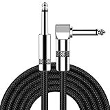
- [Low Noise High Fidelity] New bee guitar cord with 22AWG oxygen-free-copper center conductors provides enhanced signal transfer, the PVC insulation material and the high-density stranded copper double shield provide noise rejection for quiet operation
- [Ideal for Choice] New bee 1/4 straight and right angle metal connectors, ensures a secure connection in any instrument, pedal, or amp jack. L shape great fro on stage guitar stands
- [Wide Compatible] Suitable for mixer, power amplifier, microphone, effector, decoder, equalizer, electoronic organ, electric guitar, electric bass and other electronic equipments. Widely used in the installation of smart home, karaoke speaker, professional stage audio, etc
- [Easy Disassemble] Making incidental repairs quick and easy! Very nice, flexible cable
- [Dont Hesite to Order] 24 hours customer services and professional technology team are standing by
Checks and fixes
- Confirm that the patient was centered and kept within the CT field of view.
- Look for missing arms, shoulders, lateral soft tissue, or other anatomy on the CT attenuation image.
- Use the scanner’s extended-field-of-view capability when available and validated for the examination.
- Compare attenuation-corrected and non-attenuation-corrected PET. A focus created or amplified by the incomplete attenuation map should not be interpreted as a lesion without corroboration.
The 2022 hybrid-imaging guideline specifically identifies CT truncation as a reason to inspect PET images with and without attenuation correction.
CT motion, noise, and reconstruction defects
Not every artifact is a dramatic streak or a large displacement. CT motion, poor count density, and reconstruction artifacts can subtly degrade the attenuation map or the fused display. Review CT quality separately from PET quality before deciding that an uptake pattern is biologic.
CT motion
Motion may blur the body contour or create duplicated edges. If the blurred region supplies attenuation values for an adjacent PET focus, the correction can be wrong even when the PET itself is acceptable.
Noise and low-quality CT
Excessive CT noise can make the attenuation map irregular and reduce confidence in small or low-contrast findings. Determine whether the issue affects only the CT image, the attenuation map, or both.
Rank #4

- Digital Family Calendar: BSIMB digital calendar wall touch screen puts an end to your chaotic life. Designed for busy families, the digital calendar keeps you organised and in step with your family. Assign different colours and tasks to each family member to make family scheduling easier and more intuitive.
- Easy Setup and Auto-Sync: Plug in, connect to WLAN and auto-syn your electric calendar in minutes. Digital wall calendar compatible with Google calendar, iCloud calendar, Outlook calendar, Cozi calendar and Yahoo calendar. Add events directly on the device or through the ecalendar app. Electronic calendar stays current whether you're at work or traveling—no manual updates needed.
- All-In-One Home Assistant: Smart calendar builds habits and encourage independence with personalised chores. Display daily meal plan to reduce the daily ‘What's for dinner?’ stress. To-do lists keep track of everything you want. Turning chores into valuable achievements with stars and rewards, making life easier and more enjoyable.
- Manage Anytime Anywhere: BSIMB electronic calendar for family stay connected with the powerful ecalendar app. Access and manage your digital wall calendar anytime, anywhere with free features like stars & rewards, photo screensaver, chore charts, meals plans, to-do lists etc. Electronic planner paid features like Magic Import tailor advanced tools for users with special needs.
- Stay in Touch with Loved Ones: Share photos or videos from your phone to electronic wall calendar with app, instantly update your life moment, and keep in touch with your family, especially those who are far away; you can also surprise your smart calendar digital wall by sending him a wish card; it's also great for keeping tech-illiterate elders in touch with the youngsters.
Reconstruction artifacts
Unusual streaks, rings, or edge effects should be checked on the raw available series and on the attenuation map. A PET focus that follows a reconstruction defect and is absent on non-attenuation-corrected PET should be reported as potentially artifactual rather than treated as independent evidence of disease.
How to distinguish an artifact from real uptake
| Finding | Clues favoring an artifact | Best confirmation | Interpretive consequence |
|---|---|---|---|
| Motion or registration error | Organs, contours, or interfaces do not overlap; focus shifts with alignment | Review fused images in all planes and attempt registration | Localization and sometimes quantification are unreliable until alignment is corrected |
| Respiratory mismatch | Focus lies at the heart–diaphragm or lung–liver interface and follows a displaced edge | Compare respiratory state, attenuation map, and non-AC PET | May require matched respiratory acquisition or repeat imaging |
| Metal or dental hardware | Focus follows a CT streak or lies beside an implant | Inspect metal-related attenuation-map distortion and non-AC PET | Both visual interpretation and SUV can be falsely high or low |
| Iodinated contrast | Focus overlaps a dense vascular or bowel contrast column | Correlate contrast position with the attenuation map and non-AC PET | False-positive corrected activity is possible |
| Truncation | Relevant anatomy is outside or missing from the CT field of view | Check body contour, CT field, and extended-FOV reconstruction | Attenuation correction is incomplete in the affected region |
| CT noise or reconstruction defect | Irregular CT/attenuation pattern without a matching anatomic lesion | Review CT and PET quality independently, then compare PET reconstructions | Confidence in a small focus and its quantitative value is reduced |
No single test proves that uptake is real. Confidence rises when the focus persists on non-attenuation-corrected PET, aligns with a genuine CT abnormality, remains in the correct location after registration, and is not explained by a defect in the attenuation map.
Independent reader supportYour contribution helps us test, update, and keep practical guides available for everyone.When re-registration is enough—and when it is not
Re-registration is reasonable when
- The CT and PET images are individually diagnostic.
- The apparent problem is a position offset rather than severe blur, truncation, or streaking.
- The workstation can apply and display a controlled alignment correction.
- The corrected overlay places the focus in a plausible anatomic location without creating new mismatches.
Repeat acquisition deserves consideration when
- Motion has degraded the emission data, transmission data, or both.
- The attenuation map contains unrecoverable metal, contrast, or truncation defects in the region of interest.
- The suspected focus could change staging, treatment, or another major clinical decision.
- Registration produces an apparently better overlay but leaves the underlying images non-diagnostic.
Guidance does not establish one vendor-neutral numeric threshold for rescanning. The responsible choice balances the diagnostic value of a reliable answer against additional acquisition, radiation exposure, workflow, and patient burden under the local protocol.
Do these 3 things before closing this tab:
1Clear out junk files and repair common Windows errors2Fix the driver behind crashes, sound loss and screen glitches3Repair Windows errors before they cause bigger problemsHow to document an artifact in the report
Describe the suspected source, the affected region, and whether the limitation is qualitative, quantitative, or both. A useful report should make clear what was checked rather than simply labeling a focus “artifact.”
- Alignment: State whether PET and CT are aligned and identify any residual respiratory or positional mismatch.
- CT cause: Name the visible cause—metal streak, dense contrast, truncation, motion, noise, or reconstruction defect—when one is present.
- Attenuation comparison: State whether the focus persists on non-attenuation-corrected PET.
- Anatomic correlation: Note whether CT shows an independent lesion at the same location.
- Impact: Explain whether the artifact prevents reliable localization, qualitative interpretation, SUV measurement, or all three.
- Action: Recommend re-registration, review of an alternative reconstruction, repeat acquisition, or correlation with another examination only when the limitation warrants it.
A focus should be called suspicious only after these checks. If uncertainty remains, identify exactly which dataset is deficient and avoid presenting an unreliable SUV as definitive evidence.
A compact decision framework for the reading room
- Is the overlay anatomically aligned? If no, correct registration and reassess the focus.
- Is the CT itself diagnostic? If no, determine whether motion, metal, contrast, truncation, noise, or reconstruction is responsible.
- Does the attenuation map reproduce the defect? If yes, expect propagation into corrected PET.
- Does uptake remain on non-AC PET? If no, attenuation correction is implicated; correlate with CT before calling disease.
- Is there an independent CT correlate after correction? If yes, interpret the PET in that anatomic context rather than from the fused color image alone.
- Can software correction restore a trustworthy result? If no and the finding matters clinically, weigh repeat acquisition under the local protocol.
Key points to retain
- Patient misregistration is the most common source of artifacts reported for PET or SPECT/CT, while respiratory motion is the most common artifact demonstrated on PET/CT.
- Metal, iodinated contrast, and CT truncation can corrupt attenuation correction and create apparent uptake that is falsely high or falsely low.
- Non-attenuation-corrected PET is an essential safeguard whenever uptake may be caused by the CT attenuation map.
- Re-registration can fix a position offset, but it cannot repair emission or transmission data that motion has already degraded.
- There is no universal numeric rule for repeating an artifact-affected scan; the clinical importance of the finding and the quality of the available data determine the next step.
Frequently Asked Questions
Why can PET uptake disappear on non-attenuation-corrected images?
The apparent activity may have been created or amplified by CT-based attenuation correction. If it disappears on non-attenuation-corrected PET, inspect the CT and attenuation map for metal, contrast, truncation, or another correction defect before calling it disease.
Can metal make an SUV unreliable even when the PET focus looks convincing?
Yes. Metal can produce incorrect attenuation coefficients and falsely high or low corrected uptake. SUV measurements near a visibly corrupted attenuation map should not be treated as reliable without a validated correction and anatomic correlation.
What’s actually slowing this PC down?
Pick the symptom - the matching free tool is one click away.
Is a repeat PET/CT automatically required when respiratory mismatch is present?
No. If both datasets remain usable, review and registration may be sufficient. Repeat acquisition is considered when the mismatch or associated motion makes the result diagnostically important and not recoverable by correction.
What should be recorded when an artifact limits interpretation?
Document the source, the affected region, alignment status, whether the focus persists on non-attenuation-corrected PET, any CT correlate, and whether qualitative interpretation or SUV quantification is affected.
Quick Recap
Product prices and availability are accurate as of the date/time indicated and are subject to change. Any price and availability information displayed on Amazon at the time of purchase will apply.

