To measure a stimulation response, pair a defined stimulus with a readout suited to the tissue and biological question, then verify that the stimulation and recording systems worked as intended. A signal that follows stimulation is not, by timing alone, proof of a neural response: electrical artifacts, equipment noise, and the measurement chain can shape or mimic what you record.
What a stimulation-evoked response tells you
A stimulation-response experiment has three linked parts: the preparation, the stimulus delivered to it, and the measurement used to observe activity. The recorded signal reflects both the biology and this experimental chain. A change in calcium-dependent fluorescence, an extracellular nerve potential, and an fMRI signal are different observables; they should not be described as interchangeable measures of neural activity.


As an Amazon Associate I earn from qualifying purchases.
Interpret the signal in the context of what the readout actually measures. For example, the Park, Lipton, Sun, and Dadarlat protocol records fluorescence changes associated with calcium fluctuations in awake, chronically implanted mice using two-photon imaging. It offers a way to compare baseline and post-stimulation activity in that particular cortical preparation, not a universal measure of every neural response.
Free tools Windows power users keep installed
One-click scans. No signup required.
Choose a readout for the preparation and question
| Approach | What is measured | Documented context and main qualification |
|---|---|---|
| Two-photon calcium imaging | Fluorescence changes used as an indicator of calcium fluctuations associated with neural activity. | Park and colleagues’ protocol addresses electrical stimulation in awake, chronically implanted mice. Its example procedure is specific to that preparation; it does not establish universal settings or a head-to-head advantage over electrophysiology. |
| Electrophysiology | Electrical activity, including nerve activity recorded from an ex vivo mouse sciatic nerve in the cited protocol. | The Bio-protocol procedure provides practical troubleshooting for noise, stimulation artifacts, and equipment checks in its ex vivo context. ISCEV guidance addresses calibration and verification for clinical electrophysiology of vision, a separate scope. |
| Concurrent tES-fMRI | An fMRI signal recorded during low-intensity transcranial electrical stimulation. | The ContES checklist is designed for this specific combination. Its reporting and safety/noise considerations should not be treated as a complete checklist for all stimulation experiments. |
These sources do not provide a single head-to-head comparison across methods. Choose based on the biological quantity of interest, the tissue and preparation, the timing and synchronization your question requires, and the artifacts or noise relevant to the recording setup.
#1 Best Overall
Validate the response before calling it biological
- Define the preparation and observable. State the tissue or preparation, stimulation modality, and what the measurement reports. Distinguish calcium-dependent fluorescence, electrical potentials, and fMRI signals rather than using “neural activity” as if every method measured the same thing.
- Verify the stimulus and acquisition chain. Confirm that the stimulator delivers the intended stimulus and that the recording hardware and digitization operate correctly. The ex vivo sciatic-nerve protocol includes troubleshooting checks involving the stimulator, digitizer, and headstage; those examples belong to its experimental context.
- Characterize the electrode interface when electrodes are used. Electrode properties can affect stimulation and recording performance. Boehler and colleagues’ 2020 Nature Protocols tutorial proposes standardized performance tests to make neural-interface electrode comparisons more transparent. Describe the tests used and their limits rather than treating an electrode’s nominal specifications as proof of in-tissue performance.
- Look for artifacts and environmental noise. A stimulation artifact can obscure or resemble a response. The ex vivo nerve protocol notes that peristaltic pumps can introduce electrical noise or action-potential-like artifacts, and discusses checking whether a stimulation artifact matches the delivered current during troubleshooting. For concurrent tES-fMRI, consult the ContES checklist’s safety and noise-test considerations for that setup.
- Check timing and instrument characteristics. Synchronize stimulation and acquisition so that response timing can be interpreted. ISCEV’s 2023 guidance says stimulus and acquisition-system characteristics affect evoked waveform amplitude and peak time, and calls for regular verification and periodic calibration for reliable sequential monitoring and comparison. This guidance is for clinical electrophysiology of vision; do not present it as a universal calibration standard for every neural experiment.
- Assess whether the response is reproducible and analytically credible. Report the trials collected, signal quality, synchronization, and any averaging, artifact rejection, or noise assessment used. Explain how those choices affect the reported result; an apparent response should not be treated as causal evidence merely because it follows a stimulus.
Report enough detail for others to interpret the result
Give readers the information needed to understand both the biological preparation and the measurement chain. Include:
- The tissue or preparation, stimulation modality, and relevant stimulus parameters.
- The recording modality and what physical or biological quantity its signal represents.
- The electrode configuration, where applicable, and the electrode-characterization methods and limitations.
- How stimulation and acquisition were synchronized, and how the equipment and settings were verified.
- The noise and artifact controls, signal-quality assessment, trial handling, and analytical treatment.
The 2022 ContES consensus paper illustrates why explicit reporting matters within its own scope: among the 57 concurrent tES-fMRI papers it assessed, papers reported 24% to 76% of checklist items, with an average of 53% per paper. Those figures describe the papers in that study, not neural-stimulation research generally.
Rank #2
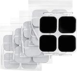
- Huge 20-Piece Set. We're always looking for new ways to bring our customers incredible value! When you shop with us, you'll receive 20 electrode pads in this jumbo pack. Each one is flexible and soft to the touch, helping you get the most from your TENS machine or EMS. They also feature gray cloth backing for durability and re-sealable packaging for a longer lifespan
- Trusted Quality. Low impedance solid electrode gel is made by upgraded industry leading adhesive, which can provide excellent self-adhesive performance and easy clean up for maximum 45 uses. Our medical grade reusable electrodes are latex-free and non-irritating to ensure users of all skin types have a safe, comfortable experience
- Universal Compatibility. Standard 2mm connector TENS electrodes pads are classic and commonly used in medical area, which makes it easier for you to find compatible TENS unit muscle stimulator & accessories
- CE, ROHS Approved. Highly ensured for optimal safety when getting fast relief through certifications, further protects you from harmful additives and consumption of prescription medication
- What You Receive: 20 x Square 2" TENS pads for TENS Unit, our 12-month warranty and technical support, and friendly customer service
Keep conclusions within the experiment’s scope
The cited procedures and guidance cover different settings: implanted electrical stimulation with two-photon calcium imaging in mice; ex vivo mouse sciatic-nerve electrophysiology; concurrent non-invasive tES-fMRI; neural-interface electrode testing; and clinical vision electrophysiology. Their methods are not one interchangeable protocol. Use the procedure or checklist suited to your preparation, and distinguish directly measured observations from interpretations about underlying neural activity.
Recommended Free Tools
Quick Recap
Best Value
Rank #4

- Excellent Self-Adhensive: The low impedance solid electrode gel is made by an industry-leading adhesive, which provides excellent self-adhesive performance for a maximum of 35 uses. Our medical-grade reusable pads are bio-compatible,latex-free, and non-irritating to ensure a safe, comfortable experience.
- Universal Compatibility: Compatible with 3.5mm snap-on connectors, TENS unit replacement pads are suitable for all muscle stimulators utilising this connectors, including TENS from NURSAL, Belifu, NueMedics, TECHCARE, MEDVICE, and TECBEAN. (Note: Incompatible with: a. Non-3.5mm connectors b. Non-snap-on connectors c. Magnetic connectors )
- Premium Quality: We're always looking for new ways to bring our customers incredible value! Each pad is flexible and soft to the touch, helping you get the most from your TENS or EMS unit. They also feature durable cloth backing and come with re-sealable packaging for a longer lifespan.
- Safety Certification: When using NURSAL TENS pads, you get fast pain relief, and are further protected by not having to ingest potentially harmful additives or consume prescription medication. They are good sticky, quick to grab, easy to peel, and dry-out resisting.
- 20 Pieces Valued Pads: Totally 20 pieces of pads, coming in small*4 (1.2''×1.6''), medium*12 (1.6''×1.8''), and large*4 (2''×4'') sizes, the electrodes can target different massage locations on the body for a better massage experience.
Rank #3
Product prices and availability are accurate as of the date/time indicated and are subject to change. Any price and availability information displayed on Amazon at the time of purchase will apply.




