The Tool Desk
Outbyte PC Repair FREEClear out junk files and repair common Windows errorsFree Scan →Outbyte Driver Updater FREEFix the driver behind crashes, sound loss and screen glitchesFind Drivers →For a microscope image to be reproducible, preserve more than the pixels: keep the original acquisition data together with sample context, the microscope configuration, settings that shaped the image, image dimensions and scale, relevant calibration records, and a traceable account of processing. Record the details that matter to the experiment rather than treating every metadata field as universally required.
What a reproducible microscope image record contains
An image by itself rarely describes how it was made. Another researcher needs enough context to interpret the image and, where appropriate, repeat the acquisition: what sample was imaged, which instrument and configuration were used, how the image was acquired, and how the data were subsequently processed or presented.
As an Amazon Associate I earn from qualifying purchases.
The Open Microscopy Environment (OME) describes a compliant file as containing enough information to authoritatively describe an imaging experiment so that someone with the same sample and microscope could reproduce the recorded data. That is a target for meaningful documentation, not a guarantee that every microscope embeds every necessary field or that every field applies to every modality.
Do these 3 things before closing this tab:
1Clear out junk files and repair common Windows errors2Scan for outdated or missing drivers - takes under a minute3Repair Windows errors before they cause bigger problems- Sample context: a stable sample identifier and the preparation, treatment, or protocol information needed to interpret it.
- Instrument context: the microscope or facility identifier, objective, and relevant optical, illumination, and detector components.
- Acquisition record: the date and settings that materially affect the data, such as modality, channels, exposure or dwell time, illumination or laser settings, and scan or time-series parameters where applicable.
- Image structure: dimensions, data type, axes or dimension order, physical pixel size, and relevant z, time, position, and channel information.
- Measurement context: calibration and quality-control information relevant to the quantitative claims being made.
- Data lineage: original files, derived images, processing steps, analysis, and figure presentation information connected to one another.
Choose the right level of documentation
Documentation should scale with the experiment’s complexity and purpose. The 4DN-BINA-OME specifications, developed through community work that includes QUAREP-LiMi, use five tiers to organize increasing imaging and analytical complexity. They cover microscope hardware, acquisition settings, and calibration procedures; researchers can use the tier-appropriate subset instead of filling out an exhaustive list for every image.
#1 Best Overall

- 【WiFi & USB Microscope】This is a wireless handheld digital microscope that has been designed to work with your mobile Android or iOS device (open your device’s WiFi to connect to the microscope's WiFi hotspot), also compatible with Windows or Mac computers (via USB cable)
- 【8 Adjustable LED Lights】The microscope camera has 8 adjustable LED lights that provide excellent detail and optimal clarity, allowing you to capture digital images at 1920x1080 resolution. 1080P HD picture quality for the smartphone, 720P for the computer
- 【One-Button Photo/Video Capture】Simply tap the camera button on the device or trigger via the app to instantly take a photo or record a video. This WiFi handheld digital microscope, equipped with a 2MP HD CMOS sensor, transmits the captured images or videos directly to your smartphone
- 【Portable Microscope 】Lightweight and small size are convenient for taking them with you everywhere. Easy to operate, allows you to take it on your trips for children to study plants, minerals, insects, or have fun outdoor activities. This electronic microscope is more of a fixed focus magnifying glass, not a traditional microscope, Not suitable for professional serious biologists!
- 【Optimal Focal Length Range】3-60 mm. To ensure image sharpness, please ensure that the distance between the microscope lens and the object being observed is maintained within the range of 3-60 mm.
For a routine qualitative image, record enough to identify the sample, instrument, objective, acquisition mode, and relevant settings. Quantitative, confocal, live-cell, multiphoton, super-resolution, or custom-instrument work can require additional acquisition and calibration detail. The exact fields depend on the modality and the conclusions the experiment is meant to support. Check the current specification and facility procedures because the community guidance continues to develop.
Before acquisition: connect the sample to the instrument
- Set the documentation scope. Identify the experimental question and decide which tier-appropriate details another person would need to assess or repeat the acquisition.
- Assign a durable sample identifier. Record preparation, treatment, and protocol context in the laboratory record, and make the connection between that record and the image dataset unambiguous. Use the sample fields required by the experiment and any relevant repository.
- Identify the microscope and configuration. Record the instrument make and model or facility identifier. Note relevant components, which may include the objective and immersion medium, optical path, filters, illumination source, and detector.
- Check calibration needs. If the interpretation depends on spatial scale or another instrument property, make the applicable calibration and performance information available. Use the facility’s approved procedures; community guidance addresses optical, excitation, wavelength, mechanical, and detector calibration.
During acquisition: capture settings that affect the result
Preserve the acquisition date and time and the settings that materially shape the recorded image. OME documentation illustrates relevant fields including image dimensions and data type, physical pixel sizes, channel names, excitation and emission wavelengths, exposure, detector settings, positions, and time intervals. Which fields apply depends on the experiment.
Rank #2

- 4.3'' Crisp Display: With 720P HD digital imaging, the LCD screen displays real-time clear images and good quality videos in full lighted view with 8 LED fill lights; A large screen enhances ergonomics and eliminates eye and neck strain
- Powerful Magnification: This microscope for adults allows you to zoom in and see the incredible details at a great magnification. The actual magnification differs due to the screen size, the distance between the camera and object
- Microscope to PC Larger View: Connect this electronic microscope effortlessly to Windows or MacBook for viewing on a larger scale, and share live images with multiple viewers; Also easily saving and organizing your images or videos for better analysis
- Enhanced Viewing with LED Lighting: This handheld microscope features 8 adjustable LED lights that provide optimal illumination, ensuring clear and detailed observations even in low-light conditions, and improving image quality
- Widely Used, Great Gift: Suitable for circuit board inspection, coins, jewelry, and stamps, watch, clock repair, skin detection, kids STEM education, textile industrial, QC inspection and more
- For fluorescence imaging, record the illumination and acquisition mode and the settings for each channel, including relevant excitation and emission information.
- For confocal or multiphoton imaging, include applicable modality-specific parameters, such as pinhole or laser-related settings.
- For multidimensional datasets, preserve the z, time, and position information and intervals needed to interpret the series.
- For quantitative work, retain physical pixel size and other applicable calibration context alongside the values used for interpretation.
Do not mechanically fill every possible field: some settings are relevant only to particular modalities. Conversely, do not assume the instrument’s embedded metadata are a complete record. Manufacturer metadata varies, and published microscopy work has described cases where embedded information does not meet minimal information criteria. Keep a contemporaneous record of important settings that the system does not save.
Quick wins for a faster PC:
Scan for outdated or missing drivers - takes under a minuteDriver Scan →Repair Windows errors before they cause bigger problemsFix Now →Fix the driver behind crashes, sound loss and screen glitchesFind Drivers →After acquisition: preserve originals and add structured metadata
Keep the original acquisition files. If you create stitched, deconvolved, projected, analyzed, or figure-ready images, retain those as derived data and document how they relate to the originals. Record material software and version information, transformations, analysis steps, and display adjustments relevant to interpreting the result.
Rank #3
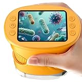
- 【1000X Magnification & 2MP Camera】Real 1000X optical+digital magnification with manual focus control - seeing actual plant cells and insect anatomy with crystal-clear 2MP resolution. Unlike toy microscopes, our precision-engineered manual focus system delivers lab-quality imaging, while the anti-shake design keeps specimens perfectly visible even in excited little hands. STEM-certified for real science learning, this is the tool that turns backyard exploration into profound discovery.
- 【ABS Plastic Material, CE/FCC/CPC Safety Certified】Kid-Friendly Design,Made from eco-friendly ABS plastic, with CE/FCC/CPC triple safety standards, odorless and smooth with no rough edges, suitable for children aged over 3 years.oversized focus dials for uncoordinated fingers, shock-absorbent rubber casing that survives playground drops, and a featherlight 200g body. Tests proved 92% of kids could operate it alone within minutes - no frustrating setup, just instant scientific empowerment.
- 【Anti-Shake & 8 Adjustable LEDs & 600mAh battery】Our exclusive 8-LED system with 3 adjustable brightness modes (Off/Medium/High) reveals hidden details in any light. The diffuser prevents glare while illuminating specimens perfectly - whether studying fireflies at dusk or creating a midnight "bedroom lab." The 600mAh battery lasts through 2 hours of discovery, stays powered via USB or portable charger for unlimited adventures.and the included carry lanyard makes it perfect for on-the-go exploration—whether in the backyard or at a science museum.
- 【Photo & Video Storage: Capture, Save, & Share】Every "Whoa!" moment gets preserved with 32M storage for 200+ high-res photos or AVI videos,perfect for documenting science experiments, nature explorations, or classroom projects. Easily export content to any Windows (XP/Vista/7/8/10/11) or Mac (11.0+) system via USB, allowing kids to create digital portfolios, share discoveries with teachers, or compile family-friendly science albums. Perfect for school presentations, science fairs, or creating a growing "research portfolio" of their explorations
- 【Gift-Ready Packaging &1-year warranty】Comes in gift box —ideal for birthdays, Christmas, or "learning milestone" presents. Beyond a toy, it fosters patience and curiosity through manual focusing for kids.Backed by a 12-month worry-free warranty, it creates those priceless moments when a child first sees the magic in science - with 2000+ parents reporting it's the rare gift that gets used daily for months. Not just a toy, but a passport to lifelong curiosity.
Associate a structured settings record with both the image dataset and the microscope configuration. Micro-Meta App is one open-source approach described in a 2021 Nature Methods paper: it can import available OME-compatible metadata through Bio-Formats, prompt for missing acquisition details, and create interoperable Microscope.JSON and Settings.JSON records. The paper reports use at 16 partnering core facilities and more uniform metadata fields than a Bio-Formats-only baseline in that usability case study. That result describes those facilities, not a guaranteed outcome for every instrument or lab; check current software documentation before relying on particular features.
Independent reader supportYour contribution helps us test, update, and keep practical guides available for everyone.Choose a file format and sharing arrangement that fit the workflow
There is no single correct format for every microscope, analysis pipeline, and repository. Choose an arrangement that retains interpretable image structure and metadata and works across the acquisition system, downstream tools, collaborators, and target repository.
Rank #4

- Higher Frame Rate: 5MP USB digital camera with live image speed of up to 30 frames per second under maximum resolution 2592x1944, keeps the motion smooth with details
- Higher Sensitivity: Sony Back-illuminated sensor guarantees higher sensitivity and signal-to-noise ratio, delivering high-speed live display, livestream videos and quality photos
- USB Software Installation Disk: Included USB flash disk contains installation software, which is convenient to use compared with CD. Software is also available for download online
- Compatible with Various Microscopes: Fits 23.2mm, 30mm and 30.5mm microscope eyepiece tubes with the 30mm and 30.5mm two additional adapters included
- 5 Megapixel Digital Eyepiece Camera: Designed for compound and stereo microscopes, allowing lecturers, instructors, and clinicians to share images with large audiences
- OME-TIFF: an OME-related option for retaining image data with structured metadata. Confirm that your acquisition and analysis tools preserve the fields you need when exporting or converting.
- OME-Zarr: combines Zarr storage for scientific arrays with an OME metadata model for bioimaging. It may suit large or cloud-accessible datasets when the lab’s tools and repository support it.
- Vendor-native or other formats: may be needed within an acquisition ecosystem, but check whether the metadata remain readable in the software your collaborators or repository use. Preserve the original when converting.
OME-Zarr and related specifications evolve. Before converting or depositing data, check the specification version and features supported by the actual acquisition, analysis, and repository systems. Include a stable dataset identifier or repository record and any access or reuse conditions required for sharing.
Recommended Free Tools
Document processing and prepare publication figures transparently
A displayed figure is not a substitute for the underlying image data and acquisition record. Keep the figure traceable to the source files, and report enough about the analysis and presentation for readers to understand how the displayed result was produced.
- Record material image-analysis steps and the software and version where they affect interpretation.
- Document transformations and display-range or contrast adjustments relevant to the figure.
- Use clear annotation and color choices, and explain channel or color conventions where needed.
- Provide the data-availability information and links or identifiers required by the journal and repository.
- Check the journal’s and repository’s current instructions before submission; requirements are not universal.
A community-developed 2023 preprint proposes publication checklists addressing formatting and annotation, color selection, data availability, and image-analysis workflow reporting. Treat that as useful community guidance alongside the current rules of the journal receiving the manuscript.
Adaptable microscope-image documentation checklist
Use this as a planning aid, not a requirement to record every field for every experiment. Select the applicable details based on modality, experimental purpose, quantitative needs, and repository rules.
Quick Recap
- Sample and context: sample identifier, preparation or treatment, labels or channels, and link to the notebook or protocol.
- Microscope configuration: instrument identifier and model; objective and immersion information; illumination path and relevant optical components; detector identity and relevant settings.
- Acquisition: date and time; modality and mode; channels and relevant excitation or emission wavelengths; exposure or dwell settings; illumination or laser power where applicable; gain, binning, scan or frame settings; z, time, and position information where applicable.
- Image structure: axes and dimension order, pixel type, dimensions, physical pixel size, z spacing where applicable, channel names, and timing or position metadata for multidimensional experiments.
- Calibration and quality control: procedure and date, plus relevant optical, mechanical, excitation, wavelength, or detector measurements for the claims being made.
- Processing and presentation: material software and version, transformation and analysis steps, relevant display adjustments, annotation and color conventions, and the relationship between processed views and originals.
- Sharing: stable dataset identifier or repository record, format and specification version, associated structured metadata, and access or reuse conditions.
Product prices and availability are accurate as of the date/time indicated and are subject to change. Any price and availability information displayed on Amazon at the time of purchase will apply.




