Brain imaging is how researchers collect measurements; brain mapping is the work of turning those measurements into an interpretable map. MRI can show anatomy, fMRI can infer activity from blood-oxygenation changes, and diffusion MRI can estimate large-scale white-matter pathways. Connectomics analyzes connections at a declared scale, from networks linking major regions to neuron- or synapse-level maps in small pieces of tissue. These terms overlap, but they are not interchangeable—and none of them means that a routine scan has produced a complete, cell-by-cell wiring diagram of a living human brain.
What’s the difference between brain mapping and brain imaging?
Brain imaging refers to measurement techniques that acquire data from the brain. Brain mapping is the scientific goal or analytical product: a map that relates anatomy, physiology, activity, or connectivity to locations in the brain.

As an Amazon Associate I earn from qualifying purchases.
The National Library of Medicine’s Brain Mapping MeSH descriptor (data labeled 2025) uses a narrower definition: “Imaging techniques used to colocalize sites of brain functions or physiological activity with brain structures.” In everyday research writing, however, mapping can also include structural atlases, connectivity matrices, and other analyses that combine imaging with behavioral or demographic data.
A useful way to keep the distinction straight is to ask two questions:
#1 Best Overall

- [Ultra-Lightweight Ear Buds Designed for Small Ears] Each earbud weighs only 3.7g and features a compact, ergonomic in-ear design made especially for small ears. Secure, low-profile, and comfortable for workouts, all-day wear or overnight sleeping.The ultra-slim profile sits flush in your ear with no hard edges poking or rubbing, so you can even sleep on your side without discomfort.
- [Immersive Stereo Sound with TOZO OrigX Technology] TOZO OrigX tuning delivers clear vocals, balanced mids, and natural stereo sound for music, podcasts, and videos.
- [Long Battery Life for Daily Use] Get up to 7 hours of playtime on a single charge, with up to 32 hours total using the charging case—ideal for workdays, commuting, and extended listening sessions.
- [Bluetooth 5.3 & Stable Connection] Bluetooth 5.3 provides fast pairing, stable wireless performance, and reduced dropouts as you move around home or office.
- [Deep Bass with Clear Vocals] High-performance drivers produce punchy bass while keeping vocals clean and detailed for everyday listening.
- What signal was measured? Magnetic-resonance contrast, blood-oxygenation changes, water diffusion, or another signal.
- What map was inferred? Anatomy, task-related activity, a functional network, estimated pathways, or a connectome at a specified scale.
The same MRI dataset can contribute to several maps, while a “map” may combine more than one imaging method. The map is therefore an interpretation of measurements, not a photograph of every process taking place in the brain.
What structural MRI actually shows
Structural MRI uses magnetic-resonance contrasts that vary with tissue properties. The resulting images provide a high-detail reference for anatomy: the shape and volume of brain structures, tissue boundaries, and other visible features. This is the MRI most people mean when they say a scan “shows the brain.”
Structural information is essential for mapping because functional or connectivity results need to be located in an anatomical space. Researchers can align measurements from different people to a common coordinate system, identify regions of interest, and compare tissue characteristics across groups.
There is an important boundary, however. Anatomy alone does not establish moment-to-moment neural activity, and it does not reveal every connection between regions. A structural image can provide the scaffold on which other maps are placed; it is not itself a complete activity or wiring map.
What an anatomical map can and cannot answer
- It can help answer: Where are major structures? How large or thick is a region? How does anatomy differ between observations?
- It cannot answer by itself: Which neurons are firing right now, whether two regions are functionally coordinated, or whether every axon between them has been traced.
How fMRI infers brain activity
Functional MRI (fMRI) measures changes related to blood flow and oxygen consumption. Those hemodynamic changes are associated with neural activity, so researchers use them as an indirect marker. fMRI is not a direct recording of neurons firing and should not be described as watching individual neurons in real time.
Task-based fMRI
In task-based fMRI, a participant performs an instructed task—such as viewing images, moving a finger, or recalling information—while the scanner records signal changes. Analysts compare activity-related signals during different conditions or against a baseline, then relate differences to behavior or task performance.
The resulting map answers a constrained question: which locations show different hemodynamic responses under the tested conditions? It does not mean that a region is dedicated exclusively to the task, nor that the measured response is a direct count of neural spikes.
Free tools Windows power users keep installed
One-click scans. No signup required.
Resting-state fMRI
Resting-state fMRI collects data while a participant is not performing a structured task. Researchers examine correlated spontaneous fluctuations in the fMRI signal across areas. Groups of regions whose signals fluctuate together can be represented as functional networks.
Rank #2

- 【IP68 Waterproof Multifunctinal Smart Watch】Torjalph fitness activity tracker smart watch is IP68 waterproof technology ,you can wear it when swimming,handwashing,it's rain-proof which means that you perform multiple exercise modes outside without worrying about sweat or rain corroding your fitness smart watch,and this watch equipped with a 230 mAh large capacity battery magnetic charging ,and this sports activity tracker watch can be used 5-7 days If fully charged.
- 【Larger 1.83'' Full Screen Watch & Multi-dial Selection】1.83 inch full HD color touch screen smart watch for men / women provides excellent 240*284 resolution image quality ,This Bluetooth smart watch offers a better touch experience and remarkable immersion and ,more than 200 watch faces for you to choose from App"Da Fit ", you can change the dial style according to your mood to wear keeping the world on your wrist
- 【Accurate Activity Tracker Smart Watch 】sports watch activity tracker uses sweat and dust resistant breathable silicone strap, our Fitness Tracker Smart Watch for Android iOS Phones supports 12 sports modes,walking,running,bicycle,swimming,badminton,basketball,football,rope skipping. This bluetooth calls smart watch for men women is lightweight and stylish, bringing more convenient and relaxing sports experience while also providing more protection.
- 【Your 24 Hour Personal Daily Health Monitor】Fitness watch for women men full touch screen IP67 waterproof bluetooth smartwatch for Android iOS Phones with heart rate blood pressure monitor multifunction Andriod smart watch with steps counter sleep monitor sports fitness watch, monitoring blood oxygen saturation allows you to understand your physical condition in real time to track fitness progress ,to know your health status in time.
- 【More Functions Bluetooth Call Smart Watch】This fitness activity smart watch full screen for iPhone and Android phones includes a bunch of new features including:The bluetooth smart watch can hang up the phone, receive and display WhatsAPP /Text /Facebook /Twitter /Messenger reminder.Support sedentary reminder, alarm clock, stopwatch, timer,weather information,music control, remote camera control compatible with Android 5.0 and above or iOS 9.0 and above smartphones.
These correlations are useful for studying large-scale organization, but correlation is not proof of a direct anatomical connection. Two areas can show related fluctuations because of indirect pathways, shared inputs, global physiological effects, or analysis choices. NIH guidance emphasizes that resting-state functional networks correspond to anatomical connections only imperfectly.
Diffusion MRI and estimated structural pathways
Diffusion-weighted MRI uses the directionality of water diffusion, especially in white matter, to estimate the trajectories of long-distance pathways. Analyses can produce tractography or a structural-connectivity matrix describing likely links among larger regions.
The word estimate matters. Diffusion measurements are indirect, and the reconstructed pathways depend on the data, modeling assumptions, resolution, and tracking method. Crossing, branching, or tightly packed fibers can make interpretation difficult. A tractography line is not a photograph of an individual axon, and a diffusion-derived map should not be presented as a complete inventory of the brain’s wiring.
Structural versus functional connectivity
Structural (anatomical) connectivity refers to physical pathways, estimated at the macroscale with methods such as diffusion MRI. Functional connectivity refers to statistical relationships between activity-related signals, often measured with resting-state fMRI. The two can be related without being identical.
A pair of regions may be functionally correlated without a direct physical fiber between them, and a physical pathway may not produce a strong correlation during a particular state. The method and the question determine what a connectivity result means.
What “connectomics” means
Connectomics is the study and mapping of neural connections. A connectome is not one universal object: it is a representation of connections made at a stated spatial scale, using a stated modality and analysis.
Macro-connectomes
Macro-connectomes describe connections among larger brain regions across much or all of the brain. Current human connectomic research relies heavily on MRI-based methods—especially diffusion MRI for estimated structural pathways and fMRI for functional networks. These maps are appropriate for questions about systems-level organization, communication among regions, and variation across people or conditions.
Quick wins for a faster PC:
Fix the driver behind crashes, sound loss and screen glitchesFind Drivers →Clear out junk files and repair common Windows errorsFree Scan →Scan for outdated or missing drivers - takes under a minuteDriver Scan →Meso-connectomes
Meso-connectomes operate at an intermediate scale, between whole-region networks and individual-cell maps. They may resolve finer subdivisions or local circuits than a typical whole-brain MRI analysis, but they still do not amount to a neuron-by-neuron inventory of a human brain.
Rank #3
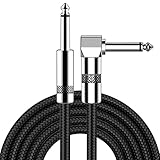
- [Low Noise High Fidelity] New bee guitar cord with 22AWG oxygen-free-copper center conductors provides enhanced signal transfer, the PVC insulation material and the high-density stranded copper double shield provide noise rejection for quiet operation
- [Ideal for Choice] New bee 1/4 straight and right angle metal connectors, ensures a secure connection in any instrument, pedal, or amp jack. L shape great fro on stage guitar stands
- [Wide Compatible] Suitable for mixer, power amplifier, microphone, effector, decoder, equalizer, electoronic organ, electric guitar, electric bass and other electronic equipments. Widely used in the installation of smart home, karaoke speaker, professional stage audio, etc
- [Easy Disassemble] Making incidental repairs quick and easy! Very nice, flexible cable
- [Dont Hesite to Order] 24 hours customer services and professional technology team are standing by
Micro-connectomes
Micro-connectomes aspire to map neurons, synapses, or very small circuits in restricted tissue. They generally require ultrastructural methods and experimental tissue rather than routine scans of living people. A microscopic map of a small sample can be extraordinarily detailed while covering only a tiny volume; it should not be conflated with a whole-human connectome.
Whenever you encounter the word “connectome,” look for three qualifiers: the scale (macro, meso, or micro), the connection type (structural or functional), and the measurement method. Without those details, the term can sound more complete than the evidence warrants.
MRI, fMRI, diffusion MRI, and connectomics compared
| Approach | Signal or basis | Typical mapping contribution | Important limitation |
|---|---|---|---|
| Structural MRI | Magnetic-resonance contrast from tissue properties | Brain anatomy and a structural reference space | Anatomy alone does not establish moment-to-moment activity or all connections. |
| Task fMRI | Changes in blood flow and oxygen consumption associated with neural activity | Activity-related differences during tasks and relationships with behavior | The hemodynamic signal is an indirect proxy, not a direct neuronal recording. |
| Resting-state fMRI | Correlated spontaneous fluctuations in fMRI signals | Functional networks inferred across dispersed areas | Functional correlations correspond to anatomical connections imperfectly. |
| Diffusion MRI | Directional water diffusion in white matter | Estimated long-distance pathways and macroscale structural-connectivity analyses | Pathways are inferred from diffusion measurements; the result is not a complete wiring inventory. |
| Connectomics | Analysis or mapping of connections at a declared resolution | A connectivity representation, structural or functional | Meaning depends on scale, modality, and whether the map concerns living humans or experimental tissue. |
How a brain map is built from measurements
Although projects differ, a typical MRI-based mapping workflow has several linked stages:
Recommended Free Tools
- Define the question and scale. Decide whether the goal is anatomy, task-related activity, spontaneous network organization, or estimated pathways. State whether the analysis is whole-brain, regional, or microscopic.
- Acquire the relevant data. Structural MRI supplies anatomy; task or resting-state fMRI supplies activity-related signals; diffusion MRI supplies direction-sensitive measurements used for pathway estimates.
- Preprocess and align the data. Researchers correct for motion and other artifacts, register scans to anatomy, and place observations in a common coordinate system when group comparisons are needed.
- Extract measurements. This may mean tissue volumes, task-condition contrasts, correlations between time series, or diffusion-based estimates of links between regions.
- Represent the result as a map or network. A connectivity matrix can be visualized as a graph, while activity and anatomy can be displayed on a surface or in three-dimensional coordinates.
- Check uncertainty and interpretation. The final map must be read in light of signal limitations, model assumptions, motion, physiological noise, and the distinction between correlation and physical connection.
This pipeline explains why “the brain map” is not a single standardized product. Different choices can produce different maps from the same underlying scans.
Why resolution and directness matter
Two maps can both be called high-resolution while resolving different dimensions. Spatial resolution concerns how finely locations are separated; temporal resolution concerns how quickly changes can be tracked. A method can be relatively detailed in one dimension and limited in the other.
Directness is another axis. Structural MRI measures tissue-dependent signal directly enough to depict anatomy, but interpreting a pathway from diffusion or activity from fMRI adds inference. The farther a conclusion is from the measured signal, the more important it is to report assumptions and uncertainty.
- Measured signal: what the scanner physically records.
- Derived feature: a calculated value such as a correlation or diffusion direction.
- Scientific interpretation: a claim about activity, communication, or anatomy based on that feature.
Keeping these levels separate prevents overstatement—for example, turning a correlated fMRI pattern into a claim of direct wiring or turning a tractography estimate into proof of an individual axon.
PC Slower Than It Used to Be?
A free scan shows the junk files, broken settings and background clutter dragging Windows down - then fixes them in one click.Free scan · Windows 10 & 11Outdated Drivers Are Slowing You Down
One free scan finds every outdated or missing driver and matches the right update for your exact hardware.Free scan · exact hardware matchThe Human Connectome Project: important, but not a complete cellular map
The Human Connectome Project (HCP) was a major research initiative aimed at acquiring and sharing data on structural and functional connectivity, together with behavioral and demographic information. NIMH describes an NIH Blueprint award of $40 million over five years beginning in 2010. Its history separately dates the project’s launch to 2009; those dates refer to different milestones and should not be collapsed into “launched in 2010.”
Rank #4

- Digital Family Calendar: BSIMB digital calendar wall touch screen puts an end to your chaotic life. Designed for busy families, the digital calendar keeps you organised and in step with your family. Assign different colours and tasks to each family member to make family scheduling easier and more intuitive.
- Easy Setup and Auto-Sync: Plug in, connect to WLAN and auto-syn your electric calendar in minutes. Digital wall calendar compatible with Google calendar, iCloud calendar, Outlook calendar, Cozi calendar and Yahoo calendar. Add events directly on the device or through the ecalendar app. Electronic calendar stays current whether you're at work or traveling—no manual updates needed.
- All-In-One Home Assistant: Smart calendar builds habits and encourage independence with personalised chores. Display daily meal plan to reduce the daily ‘What's for dinner?’ stress. To-do lists keep track of everything you want. Turning chores into valuable achievements with stars and rewards, making life easier and more enjoyable.
- Manage Anytime Anywhere: BSIMB electronic calendar for family stay connected with the powerful ecalendar app. Access and manage your digital wall calendar anytime, anywhere with free features like stars & rewards, photo screensaver, chore charts, meals plans, to-do lists etc. Electronic planner paid features like Magic Import tailor advanced tools for users with special needs.
- Stay in Touch with Loved Ones: Share photos or videos from your phone to electronic wall calendar with app, instantly update your life moment, and keep in touch with your family, especially those who are far away; you can also surprise your smart calendar digital wall by sending him a wish card; it's also great for keeping tech-illiterate elders in touch with the youngsters.
The project advanced macroscale human connectomics by combining imaging technologies and large, shared datasets. It did not produce a complete cellular or synapse-by-synapse connectome of the human brain. Its significance is better understood as a foundation for measuring and comparing large-scale structural and functional organization.
Independent reader supportYour contribution helps us test, update, and keep practical guides available for everyone.What NIH’s BRAIN CONNECTS work is trying to do
NIH’s BRAIN CONNECTS program is a current research effort to build technical capabilities and analytical pipelines for mapping connections across mammalian brains. Its goals include integrating data across modalities and sharing data so that connection maps can be compared and combined.
That program reflects active work across scales; it is not evidence that a complete human connectome at cellular resolution is already available. Specialized research systems and pipelines may extend what can be measured, but they should not be confused with the specifications or capabilities of an ordinary clinical MRI examination.
Do these 3 things before closing this tab:
1Scan for outdated or missing drivers - takes under a minute2Repair Windows errors before they cause bigger problems3Fix the driver behind crashes, sound loss and screen glitchesCommon misconceptions and how to correct them
“An MRI is a brain map.”
An MRI is a set of images or measurements. It becomes a map only after researchers define what is being represented—anatomy, activity-related changes, or connections—and analyze it accordingly.
“fMRI shows neurons firing.”
fMRI detects hemodynamic changes associated with neural activity. It is an indirect measure and does not directly record individual neuronal firing.
“A functional connection is a physical connection.”
Functional connectivity is a statistical relationship in signals. It can be informative about coordinated network behavior without proving a direct anatomical link.
“Tractography traces every axon.”
Diffusion-based pathways are estimates derived from water movement. They support macroscale structural-connectivity analysis but do not constitute a complete axonal inventory.
What’s actually slowing this PC down?
Pick the symptom - the matching free tool is one click away.
“A connectome always means every synapse.”
Connectomes exist at macro, meso, and micro scales. A whole-brain human MRI connectome and a microscopic tissue map answer different questions and cover different amounts of tissue.
How to evaluate a brain-mapping claim
When reading a paper, news release, or product description, use this checklist:
- What was measured: tissue contrast, hemodynamics, water diffusion, or something else?
- Is the result structural, functional, or both?
- What spatial and temporal scale is reported?
- Does “connection” mean a physical pathway, a statistical correlation, or a modeled estimate?
- Was the work done in living humans, other animals, or fixed experimental tissue?
- What uncertainty, preprocessing choices, and confounds could affect the map?
- Is a specialized research capability being presented as if it were routine clinical imaging?
These questions let you compare methods by the question they can answer rather than by an attractive but ambiguous label.
What these maps can—and cannot—tell an individual patient
The methods described here are primarily research tools for studying organization, physiology, and connectivity. A group-level network finding or a research connectome does not, by itself, provide a diagnosis or a treatment recommendation for a particular person. Clinical interpretation requires appropriate protocols, qualified professionals, the person’s symptoms and history, and evidence validated for that clinical use.
The Tool Desk
Outbyte PC Repair FREEClear out junk files and repair common Windows errorsFree Scan →Outbyte Driver Updater FREEFix the driver behind crashes, sound loss and screen glitchesFind Drivers →The practical bottom line is precise: imaging supplies measurements; mapping organizes those measurements into a representation; connectomics focuses specifically on connections; and every resulting map must be interpreted at its stated scale and with its modality’s limitations in view.
Frequently Asked Questions
Is brain mapping a scan that a doctor orders?
Brain mapping is usually an analysis or research objective rather than one standardized scan. MRI, fMRI, and diffusion MRI can provide data for different kinds of maps, while clinical use depends on the specific protocol and indication.
Can fMRI prove that two brain regions are connected?
No. Resting-state fMRI can identify correlated fluctuations and functional networks, but correlation does not by itself prove a direct anatomical connection.
Does a human connectome map every neuron?
No. Most whole-human connectomics is macroscale and MRI-based. Neuron- or synapse-level micro-connectomes generally require ultrastructural methods applied to restricted experimental tissue.
Quick Recap
Product prices and availability are accurate as of the date/time indicated and are subject to change. Any price and availability information displayed on Amazon at the time of purchase will apply.




