The Tool Desk
Outbyte PC Repair FREERepair Windows errors before they cause bigger problemsFix Now →Outbyte Driver Updater FREEFix the driver behind crashes, sound loss and screen glitchesFind Drivers →Yes. A medical-imaging AI can assign the correct disease label while pointing to the wrong part of the image. The reason is that image-level classification (deciding what disease is present) and localization (showing where the lesion is) are different tasks. A classifier may learn any visual pattern that predicts the label, including equipment, positioning, or workflow clues, without learning the lesion’s boundaries.
A heatmap is therefore evidence about a model’s behavior, not proof that it found the pathology. To trust a highlighted region, you need testing against expert lesion annotations, robustness checks, and—when location matters—a model trained and evaluated specifically for detection or segmentation.

As an Amazon Associate I earn from qualifying purchases.
Classification and localization answer different questions
An image-level classifier returns a label or probability such as “pneumonia present.” It is rewarded for getting that decision right across its training examples. A localization system is rewarded for identifying the lesion’s position, extent, or outline. Those objectives can diverge: a classifier can use a shortcut that correlates with disease while ignoring the abnormal tissue itself.
| Task | Output | What training rewards | What a correct result means |
|---|---|---|---|
| Image classification | Label, score, or probability for the whole image | Correct disease decision | The image is associated with the condition; it does not establish where the lesion is |
| Detection or localization | Bounding box, point, or coordinates | Agreement with annotated lesion locations | The model found a region judged abnormal by the reference annotators |
| Segmentation | Pixel- or voxel-level mask | Overlap with an expert-drawn lesion mask | The predicted extent matches the annotated abnormal tissue within the test criteria |
| Saliency visualization | Importance map generated from a model’s output | Usually no direct lesion-location objective | Shows which pixels influence the output according to that method; it is not automatically a lesion map |
A classifier’s high area-under-curve or accuracy can therefore coexist with poor lesion localization. The distinction is especially important when a clinician needs to decide where to measure, biopsy, drain, or monitor an abnormality.
#1 Best Overall

- [Ultra-Lightweight Ear Buds Designed for Small Ears] Each earbud weighs only 3.7g and features a compact, ergonomic in-ear design made especially for small ears. Secure, low-profile, and comfortable for workouts, all-day wear or overnight sleeping.The ultra-slim profile sits flush in your ear with no hard edges poking or rubbing, so you can even sleep on your side without discomfort.
- [Immersive Stereo Sound with TOZO OrigX Technology] TOZO OrigX tuning delivers clear vocals, balanced mids, and natural stereo sound for music, podcasts, and videos.
- [Long Battery Life for Daily Use] Get up to 7 hours of playtime on a single charge, with up to 32 hours total using the charging case—ideal for workdays, commuting, and extended listening sessions.
- [Bluetooth 5.3 & Stable Connection] Bluetooth 5.3 provides fast pairing, stable wireless performance, and reduced dropouts as you move around home or office.
- [Deep Bass with Clear Vocals] High-performance drivers produce punchy bass while keeping vocals clean and detailed for everyday listening.
How a model learns the wrong visual cue
Correlated equipment and workflow markers
Deep-learning systems optimize statistical prediction. If a training set contains a visual cue that appears more often in positive cases, the network can use that cue even when it is not the disease itself. A 2024 review of radiology AI “shortcuts” describes pneumonia predictions associated with portable intensive-care markers and pneumothorax predictions associated with chest tubes. Those signs can be correlated with the diagnosis because of clinical workflow, but they do not identify the pathological region.
Such shortcuts may work inside the hospital or dataset where they arose and fail at another site with different machines, protocols, or patient mix. Aggregate accuracy can hide this problem because the shortcut still produces the expected label on many internal test images.
Labels that describe the whole image
If training labels say only whether a radiograph contains pneumonia, the model is not required to learn a pneumonia border. It can distribute attention over any predictive pattern. Even a technically correct label supplies no information about which pixels were medically decisive.
Quick wins for a faster PC:
Clear out junk files and repair common Windows errorsFree Scan →Scan for outdated or missing drivers - takes under a minuteDriver Scan →Rank #2

- 【IP68 Waterproof Multifunctinal Smart Watch】Torjalph fitness activity tracker smart watch is IP68 waterproof technology ,you can wear it when swimming,handwashing,it's rain-proof which means that you perform multiple exercise modes outside without worrying about sweat or rain corroding your fitness smart watch,and this watch equipped with a 230 mAh large capacity battery magnetic charging ,and this sports activity tracker watch can be used 5-7 days If fully charged.
- 【Larger 1.83'' Full Screen Watch & Multi-dial Selection】1.83 inch full HD color touch screen smart watch for men / women provides excellent 240*284 resolution image quality ,This Bluetooth smart watch offers a better touch experience and remarkable immersion and ,more than 200 watch faces for you to choose from App"Da Fit ", you can change the dial style according to your mood to wear keeping the world on your wrist
- 【Accurate Activity Tracker Smart Watch 】sports watch activity tracker uses sweat and dust resistant breathable silicone strap, our Fitness Tracker Smart Watch for Android iOS Phones supports 12 sports modes,walking,running,bicycle,swimming,badminton,basketball,football,rope skipping. This bluetooth calls smart watch for men women is lightweight and stylish, bringing more convenient and relaxing sports experience while also providing more protection.
- 【Your 24 Hour Personal Daily Health Monitor】Fitness watch for women men full touch screen IP67 waterproof bluetooth smartwatch for Android iOS Phones with heart rate blood pressure monitor multifunction Andriod smart watch with steps counter sleep monitor sports fitness watch, monitoring blood oxygen saturation allows you to understand your physical condition in real time to track fitness progress ,to know your health status in time.
- 【More Functions Bluetooth Call Smart Watch】This fitness activity smart watch full screen for iPhone and Android phones includes a bunch of new features including:The bluetooth smart watch can hang up the phone, receive and display WhatsAPP /Text /Facebook /Twitter /Messenger reminder.Support sedentary reminder, alarm clock, stopwatch, timer,weather information,music control, remote camera control compatible with Android 5.0 and above or iOS 9.0 and above smartphones.
Post-hoc maps are not human-style reasoning traces
Methods such as Grad-CAM create a visualization from a trained network and one of its outputs. They are useful for probing behavior, but they are not a direct recording of a clinician-like chain of reasoning, nor are they definitive lesion boundaries. Different saliency algorithms can produce different maps for the same prediction, and a visually smooth region can be persuasive without being causally important.
What the radiology studies found
CheXNet: strong internal labels, weak lesion finding
In a multicenter chest-radiograph study, CheXNet achieved radiologist-level internal classification results but performed poorly at finding lesions. Its Grad-CAM examples sometimes failed to cover the intended abnormality or highlighted false-positive areas. The authors wrote: “Our data revealed that CheXNet relies highly on patterns other than the true pathologic signs to make decisions, as it showed low performance in finding the lesions but achieved radiologist-level internal classification results.” Read the study’s methods and limitations in Rethinking Annotation Granularity for Overcoming Shortcuts in Deep Learning–based Radiograph Diagnosis: A Multicenter Study.
This is not a population estimate of how often AI is wrong for the right label. It is evidence that the two performance dimensions can separate in a real, multicenter radiology setting.
Rank #3
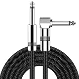
- [Low Noise High Fidelity] New bee guitar cord with 22AWG oxygen-free-copper center conductors provides enhanced signal transfer, the PVC insulation material and the high-density stranded copper double shield provide noise rejection for quiet operation
- [Ideal for Choice] New bee 1/4 straight and right angle metal connectors, ensures a secure connection in any instrument, pedal, or amp jack. L shape great fro on stage guitar stands
- [Wide Compatible] Suitable for mixer, power amplifier, microphone, effector, decoder, equalizer, electoronic organ, electric guitar, electric bass and other electronic equipments. Widely used in the installation of smart home, karaoke speaker, professional stage audio, etc
- [Easy Disassemble] Making incidental repairs quick and easy! Very nice, flexible cable
- [Dont Hesite to Order] 24 hours customer services and professional technology team are standing by
Saliency methods versus localization networks
A 2021 evaluation tested saliency approaches on two public radiology datasets and assessed localization accuracy, sensitivity to model changes, repeatability, and reproducibility. The authors reported: “All eight saliency map techniques failed at least one of the criteria and were inferior in performance compared with localization networks.” In pneumothorax segmentation, saliency methods produced area under the precision-recall curve (AUPRC) values from 0.024 to 0.224, while a U-Net localization baseline reached 0.404 on that evaluation. These are study-specific results, not universal benchmarks. See Assessing the Trustworthiness of Saliency Maps for Localizing Abnormalities in Medical Imaging.
What’s actually slowing this PC down?
Pick the symptom - the matching free tool is one click away.
Robustness can fail under tiny changes
A 2024 retrospective evaluation covered 191,229 chest radiographs and 7,022 brain MR images. It found weak saliency sensitivity to subtle input perturbations and described a commercial prototype whose saliency map could be irrelevant to changes in the model’s output. The implication is practical: if a barely perceptible image change alters the prediction but leaves the map unchanged—or changes the map without changing the prediction—the visualization is not a dependable explanation of the decision. The study is available as Revisiting the Trustworthiness of Saliency Methods in Radiology AI.
Evidence beyond chest radiographs
A separate study of automated musculoskeletal diagnoses reported that gradient-based saliency maps were not trustworthy visual explanations under its evaluation conditions. Its article record is available at Gradient-Based Saliency Maps Are Not Trustworthy Visual Explanations of Automated AI Musculoskeletal Diagnoses. Results from one modality should not be generalized automatically to every scanner, anatomy, or deployed product.
Rank #4

- Digital Family Calendar: BSIMB digital calendar wall touch screen puts an end to your chaotic life. Designed for busy families, the digital calendar keeps you organised and in step with your family. Assign different colours and tasks to each family member to make family scheduling easier and more intuitive.
- Easy Setup and Auto-Sync: Plug in, connect to WLAN and auto-syn your electric calendar in minutes. Digital wall calendar compatible with Google calendar, iCloud calendar, Outlook calendar, Cozi calendar and Yahoo calendar. Add events directly on the device or through the ecalendar app. Electronic calendar stays current whether you're at work or traveling—no manual updates needed.
- All-In-One Home Assistant: Smart calendar builds habits and encourage independence with personalised chores. Display daily meal plan to reduce the daily ‘What's for dinner?’ stress. To-do lists keep track of everything you want. Turning chores into valuable achievements with stars and rewards, making life easier and more enjoyable.
- Manage Anytime Anywhere: BSIMB electronic calendar for family stay connected with the powerful ecalendar app. Access and manage your digital wall calendar anytime, anywhere with free features like stars & rewards, photo screensaver, chore charts, meals plans, to-do lists etc. Electronic planner paid features like Magic Import tailor advanced tools for users with special needs.
- Stay in Touch with Loved Ones: Share photos or videos from your phone to electronic wall calendar with app, instantly update your life moment, and keep in touch with your family, especially those who are far away; you can also surprise your smart calendar digital wall by sending him a wish card; it's also great for keeping tech-illiterate elders in touch with the youngsters.
Why an attractive heatmap can still be misleading
Human viewers naturally treat a bright, lesion-shaped overlay as confirmation. But appearance alone does not test whether the highlighted pixels caused the prediction, whether the map is stable, or whether it would transfer to another population.
- Localization accuracy: Does the high-importance region overlap radiologist-annotated pathology at a prespecified threshold?
- Model sensitivity: Does the map change when model weights are randomized or when the output meaningfully changes?
- Repeatability: Do repeated runs under the same conditions produce similar regions?
- Reproducibility: Do independent implementations, readers, or sites obtain comparable maps?
- Perturbation robustness: Do subtle, clinically irrelevant image changes leave the explanation appropriately stable?
The 2021 saliency evaluation found that no evaluated method passed every criterion. That finding applies to the methods and datasets in that paper; it is not a declaration that every conceivable explanation technique fails on every medical task.
Outdated Drivers Are Slowing You Down
One free scan finds every outdated or missing driver and matches the right update for your exact hardware.Free scan · exact hardware matchPC Slower Than It Used to Be?
A free scan shows the junk files, broken settings and background clutter dragging Windows down - then fixes them in one click.Free scan · Windows 10 & 11How to evaluate a heatmap before trusting it
- Define the required output. If the clinical question is only whether disease is present, assess classification separately. If a user must know where the lesion is, specify detection or segmentation performance before looking at a visualization.
- Obtain an appropriate reference standard. Use lesion locations or masks annotated by qualified radiologists, ideally with an adjudication process for disagreements. Whole-image labels cannot validate a highlighted region.
- Set quantitative localization criteria in advance. Examples include intersection-over-union, lesion-level sensitivity, false-positive regions, and precision-recall measures. Report the threshold and whether the metric is calculated per image, lesion, or patient.
- Test on external data. Hold out images from another institution, scanner, protocol, or time period. A model that relies on local workflow markers may lose both classification and localization performance when those cues change.
- Run explanation robustness checks. Compare maps after controlled weight randomization, repeated runs, output-preserving transformations, and subtle input perturbations. A map that is unrelated to output changes should not be presented as causal evidence.
- Compare against a trained localization baseline. A detector or segmentation network optimized on lesion annotations provides a more direct test of “where.” The 2021 study found localization-network baselines superior to the tested saliency methods.
- Review failure cases, not only illustrative successes. Record missed lesions, false-positive highlights, device-associated regions, and cases with correct labels but incorrect locations. Examine performance by site, demographic subgroup, disease severity, and image quality.
- Document the map’s status in the interface. State whether it is a post-hoc saliency display, a detector output, or a segmentation mask. Do not label a saliency overlay as a lesion boundary unless it has been validated for that purpose.
Choosing between a classifier, localization model, and saliency method
| Approach | Best suited question | Strength | Main risk or cost |
|---|---|---|---|
| Image classifier | Is the condition likely present? | Can be trained with relatively inexpensive image-level labels | May use shortcuts and offers no guaranteed lesion location |
| Detection/localization network | Where is one or more lesions? | Optimized directly against expert locations | Requires location annotations and can miss lesions outside its training distribution |
| Segmentation network | What exact area or volume is abnormal? | Provides a measurable mask for extent and downstream planning | Pixel- or voxel-level labeling is labor-intensive; boundaries can be ambiguous |
| Post-hoc saliency map | Which image regions appear influential to an existing model? | Can be added without retraining the classifier | Reliability, stability, and lesion agreement must be demonstrated; a plausible map may still be wrong |
Do finer annotations solve the problem?
More specific supervision can help. In the multicenter chest-radiograph study, a model called CheXDet trained with finer-grained lesion annotations generally focused more effectively on pathological patterns than CheXNet in the reported examples, and some external comparisons improved. This supports annotation as a mitigation, not a guarantee.
The same study retained failure cases and did not perform as well externally as internally. Detailed boxes or masks also increase labeling time, require consistent expert protocols, and may be difficult when lesion borders are indistinct. A deployment team must weigh the expected localization benefit against that annotation and quality-control burden.
The review “Shortcuts” Causing Bias in Radiology Artificial Intelligence: Causes, Evaluation, and Mitigation emphasizes that shortcut auditing needs both domain expertise and technical skill. Data collection, labeling, evaluation design, subgroup analysis, and post-deployment monitoring all matter; improving one stage does not erase failures introduced at another.
What clinicians and patients should infer from a wrong-area highlight
A mismatched heatmap does not by itself prove that the disease label is false. It does show that the visualization cannot be used as confirmation of the lesion’s location. The label and the map should be treated as separate claims, each requiring its own validation.
For clinical use, a radiologist should verify the image and decide whether an independently validated detection or segmentation output adds value. A patient should not interpret a bright area on an AI overlay as a diagnosis, a measurement, or proof that tissue is abnormal. Questions about a particular scan belong with the qualified clinician who can review the original images, report, symptoms, and history.
Limits of current evidence
No population-level estimate establishes how often medical AI gets the disease label right while highlighting the wrong region. The cited studies cover particular radiology tasks, datasets, and evaluation designs. They demonstrate a failure mode and practical tests for it, not a universal error rate or a universally validated saliency product.
Quick Recap
Product prices and availability are accurate as of the date/time indicated and are subject to change. Any price and availability information displayed on Amazon at the time of purchase will apply.

