Quick wins for a faster PC:
Clear out junk files and repair common Windows errorsFree Scan →Scan for outdated or missing drivers - takes under a minuteDriver Scan →X-ray microscopy can look inside a battery electrode or other dense material without cutting it open. By measuring how X-rays are transmitted, absorbed, scattered, or change with energy, researchers can map particles, pores, cracks, elements, and chemical states. Tomography adds three-dimensional structure, while spectroscopic methods can show where oxidation states or compounds occur. Those images are evidence about structure and change—not a standalone certification of a cell’s safety, lifetime, or performance.
What X-ray microscopy measures
An X-ray microscope records how a beam interacts with a specimen. The transmitted intensity depends on composition, density, thickness, and photon energy; phase-sensitive arrangements can add information from the way X-rays are shifted rather than simply absorbed. Scanning instruments focus a beam on one location at a time, while full-field transmission instruments capture an area through X-ray optics. In both cases, the result is a measurement that must be interpreted with the beam energy, geometry, detector response, and sample preparation in mind.

As an Amazon Associate I earn from qualifying purchases.
X-rays penetrate much farther into many solids than visible light or electron beams. That makes it possible to inspect a representative volume of an electrode, catalyst, or fuel-cell component rather than only its exposed surface. Penetration is not unlimited: thickness, density, absorption edges, container materials, and the available photon energy determine whether a particular sample can be measured. A practical experiment therefore starts by matching the material and thickness to the instrument.
Free tools Windows power users keep installed
One-click scans. No signup required.
Full-field transmission and scanning transmission
Full-field transmission X-ray microscopy (TXM) illuminates a field of view and forms a magnified image on a detector. It is useful when a relatively large region must be recorded efficiently, including repeated views for tomography. Scanning transmission X-ray microscopy (STXM) raster-scans a focused spot and measures the signal at each position. STXM can combine high spatial detail with energy scans, allowing absorption spectra to be collected pixel by pixel. Neither approach is automatically superior: the choice depends on the required area, resolution, energy range, speed, and tolerance for dose.
#1 Best Overall

- [Ultra-Lightweight Ear Buds Designed for Small Ears] Each earbud weighs only 3.7g and features a compact, ergonomic in-ear design made especially for small ears. Secure, low-profile, and comfortable for workouts, all-day wear or overnight sleeping.The ultra-slim profile sits flush in your ear with no hard edges poking or rubbing, so you can even sleep on your side without discomfort.
- [Immersive Stereo Sound with TOZO OrigX Technology] TOZO OrigX tuning delivers clear vocals, balanced mids, and natural stereo sound for music, podcasts, and videos.
- [Long Battery Life for Daily Use] Get up to 7 hours of playtime on a single charge, with up to 32 hours total using the charging case—ideal for workdays, commuting, and extended listening sessions.
- [Bluetooth 5.3 & Stable Connection] Bluetooth 5.3 provides fast pairing, stable wireless performance, and reduced dropouts as you move around home or office.
- [Deep Bass with Clear Vocals] High-performance drivers produce punchy bass while keeping vocals clean and detailed for everyday listening.
What the main imaging modes reveal
| Mode or measurement | Primary information | Typical interpretation |
|---|---|---|
| Single-energy transmission imaging | Absorption contrast in a 2D projection | Boundaries, voids, particles, cracks, and density differences |
| Tomography | Many projections reconstructed into 3D | Particle shape, pore volume, connected pathways, tortuosity, and internal damage |
| Energy-dependent imaging | Contrast changes as photon energy is scanned | Differences in composition or chemical state when calibrated with references |
| XANES imaging | X-ray absorption near-edge spectra at image locations | Maps of oxidation state or other chemical-state signatures |
| Multi-energy tomography | 3D reconstruction at several energies | Elemental or chemical heterogeneity through a volume |
The table describes capabilities demonstrated in transmission X-ray microscopy and facility beamlines; an individual microscope may offer only some of them. Spatial and temporal resolution, field of view, energy range, and the ability to hold an operating sample vary by instrument.
From projections to a volume
Tomography rotates a specimen, acquires projections from multiple angles, and computationally reconstructs slices or a volume. A reconstructed voxel can then be segmented into active material, binder, pore space, or another phase. From that segmentation, researchers calculate quantities such as porosity, particle-size distributions, crack volume, and tortuosity—the effective complexity of pathways through a porous electrode. These are geometric measurements, not direct measurements of electrical capacity or mechanical strength.
Adding chemistry with energy
Changing the X-ray energy near an element’s absorption edge changes the contrast in a way that can carry chemical information. XANES imaging uses the shape and position of that near-edge response, compared with suitable standards or reference spectra, to distinguish chemical states. Multi-energy tomography extends the idea into three dimensions. Because spectra can be affected by thickness, orientation, self-absorption, calibration, and beam damage, a color map should not be treated as a unique chemical identification unless the analysis establishes that link.
Do these 3 things before closing this tab:
1Fix the driver behind crashes, sound loss and screen glitches2Clear out junk files and repair common Windows errors3Scan for outdated or missing drivers - takes under a minuteRank #2

- 【IP68 Waterproof Multifunctinal Smart Watch】Torjalph fitness activity tracker smart watch is IP68 waterproof technology ,you can wear it when swimming,handwashing,it's rain-proof which means that you perform multiple exercise modes outside without worrying about sweat or rain corroding your fitness smart watch,and this watch equipped with a 230 mAh large capacity battery magnetic charging ,and this sports activity tracker watch can be used 5-7 days If fully charged.
- 【Larger 1.83'' Full Screen Watch & Multi-dial Selection】1.83 inch full HD color touch screen smart watch for men / women provides excellent 240*284 resolution image quality ,This Bluetooth smart watch offers a better touch experience and remarkable immersion and ,more than 200 watch faces for you to choose from App"Da Fit ", you can change the dial style according to your mood to wear keeping the world on your wrist
- 【Accurate Activity Tracker Smart Watch 】sports watch activity tracker uses sweat and dust resistant breathable silicone strap, our Fitness Tracker Smart Watch for Android iOS Phones supports 12 sports modes,walking,running,bicycle,swimming,badminton,basketball,football,rope skipping. This bluetooth calls smart watch for men women is lightweight and stylish, bringing more convenient and relaxing sports experience while also providing more protection.
- 【Your 24 Hour Personal Daily Health Monitor】Fitness watch for women men full touch screen IP67 waterproof bluetooth smartwatch for Android iOS Phones with heart rate blood pressure monitor multifunction Andriod smart watch with steps counter sleep monitor sports fitness watch, monitoring blood oxygen saturation allows you to understand your physical condition in real time to track fitness progress ,to know your health status in time.
- 【More Functions Bluetooth Call Smart Watch】This fitness activity smart watch full screen for iPhone and Android phones includes a bunch of new features including:The bluetooth smart watch can hang up the phone, receive and display WhatsAPP /Text /Facebook /Twitter /Messenger reminder.Support sedentary reminder, alarm clock, stopwatch, timer,weather information,music control, remote camera control compatible with Android 5.0 and above or iOS 9.0 and above smartphones.
What researchers can see inside batteries
Particles, pores, and connected pathways
Electrodes are composite structures: active particles are mixed with conductive additives, binder, and pores filled by electrolyte. Three-dimensional X-ray images can show particle sizes and shapes, voids between particles, particle-to-particle contacts, and pathways through which ions and electrolyte move. SSRL describes single-energy tomography measurements of porosity and tortuosity. Those quantities help test whether an electrode’s architecture could restrict transport, but they do not by themselves establish how quickly a particular cell charges or how much energy it stores.
Cracks, voids, and structural evolution
Repeated scans can reveal particles fracturing, pores opening or closing, delamination from a current collector, and changes in the contact network. Comparing the same region before and after cycling is especially informative when registration and resolution are good enough to identify the same features. A visible crack is an observation. Concluding that it causes capacity loss requires complementary evidence linking the crack to electrical, chemical, or mechanical measurements.
Elemental and chemical heterogeneity
Energy-dependent imaging can distinguish regions that have different elemental distributions or chemical states. SSRL lists two-dimensional and three-dimensional XANES imaging for chemical states and multi-energy tomography for elemental heterogeneity. In a battery, such maps may show where a transition-metal oxidation state changes, where reaction products accumulate, or whether composition varies from the separator side to the current-collector side. Reference spectra, calibration, and a model of X-ray attenuation are needed to turn contrast into a defensible chemical assignment.
Rank #3
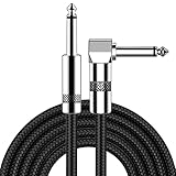
- [Low Noise High Fidelity] New bee guitar cord with 22AWG oxygen-free-copper center conductors provides enhanced signal transfer, the PVC insulation material and the high-density stranded copper double shield provide noise rejection for quiet operation
- [Ideal for Choice] New bee 1/4 straight and right angle metal connectors, ensures a secure connection in any instrument, pedal, or amp jack. L shape great fro on stage guitar stands
- [Wide Compatible] Suitable for mixer, power amplifier, microphone, effector, decoder, equalizer, electoronic organ, electric guitar, electric bass and other electronic equipments. Widely used in the installation of smart home, karaoke speaker, professional stage audio, etc
- [Easy Disassemble] Making incidental repairs quick and easy! Very nice, flexible cable
- [Dont Hesite to Order] 24 hours customer services and professional technology team are standing by
Materials while a cell operates
Ex situ imaging examines a sample removed from a cell or stopped at a selected state of charge. In situ imaging places the material in a controlled experimental environment, while operando imaging records it during electrochemical operation. Specialized cells can provide electrical connections, electrolyte, pressure, and temperature control while leaving an X-ray path. Operando data can show when a morphology or chemical change occurs during a charge or discharge rather than only what remains afterward. The cell design itself can alter thickness, pressure, heat flow, or current distribution, so results must be interpreted in that context.
Crashes, No Sound, or Screen Glitches?
Random freezes, missing sound and display glitches usually trace back to one bad driver. Find and replace yours safely.Free scan · under a minuteWindows Errors? Fix Them Before They Spread
Repair common Windows errors and clear accumulated junk for a smoother, more stable PC - no reinstall needed.Free scan · no reinstallWhy tomography is useful for degradation and failure questions
Battery-tomography reviews, including the 2017 Annual Review of Materials Research article by Pietsch and Wood, describe applications spanning structure, composition, operation, and degradation. Researchers use those observations to test mechanisms such as loss of active material, pore blockage, particle fracture, or nonuniform reaction. The method is valuable because it is largely nondestructive at the scale of the measured region and can connect a three-dimensional feature to its neighbors.
It is not a pass/fail test for a commercial cell. A scan samples a finite volume, often at a selected resolution and state of charge. A small region may miss rare defects or gradients elsewhere in a heterogeneous electrode. A proposed failure mechanism therefore needs sampling across relevant locations and, where possible, corroboration from electrochemical, spectroscopic, mechanical, or post-mortem measurements.
Rank #4

- Digital Family Calendar: BSIMB digital calendar wall touch screen puts an end to your chaotic life. Designed for busy families, the digital calendar keeps you organised and in step with your family. Assign different colours and tasks to each family member to make family scheduling easier and more intuitive.
- Easy Setup and Auto-Sync: Plug in, connect to WLAN and auto-syn your electric calendar in minutes. Digital wall calendar compatible with Google calendar, iCloud calendar, Outlook calendar, Cozi calendar and Yahoo calendar. Add events directly on the device or through the ecalendar app. Electronic calendar stays current whether you're at work or traveling—no manual updates needed.
- All-In-One Home Assistant: Smart calendar builds habits and encourage independence with personalised chores. Display daily meal plan to reduce the daily ‘What's for dinner?’ stress. To-do lists keep track of everything you want. Turning chores into valuable achievements with stars and rewards, making life easier and more enjoyable.
- Manage Anytime Anywhere: BSIMB electronic calendar for family stay connected with the powerful ecalendar app. Access and manage your digital wall calendar anytime, anywhere with free features like stars & rewards, photo screensaver, chore charts, meals plans, to-do lists etc. Electronic planner paid features like Magic Import tailor advanced tools for users with special needs.
- Stay in Touch with Loved Ones: Share photos or videos from your phone to electronic wall calendar with app, instantly update your life moment, and keep in touch with your family, especially those who are far away; you can also surprise your smart calendar digital wall by sending him a wish card; it's also great for keeping tech-illiterate elders in touch with the youngsters.
A practical workflow for an X-ray microscopy study
- Define the decision. State whether the goal is geometry, a chemical-state map, a three-dimensional elemental distribution, or a change during cycling. “Image the battery” is too broad to determine an instrument or sample design.
- Choose the specimen and thickness. Decide whether to scan a coin-cell component, a harvested electrode, a particle, or a custom operando cell. Estimate X-ray transmission through the active material, electrolyte, windows, and holders at the intended energy.
- Select contrast and dimensionality. Use a 2D map for a fast overview, tomography for connected structure, and energy scans or XANES when chemical states are central. If the feature of interest is lithium or hydrogen-rich material, evaluate whether neutron imaging could add sensitivity that X-rays lack.
- Plan controls and references. Include blank measurements, standards or reference spectra for chemical analysis, and repeated scans to assess drift. For cycling studies, define the current, voltage window, temperature, pressure, and scan points before collecting data.
- Acquire with dose and time in mind. Set exposure, angular sampling, and energy points so that the signal is adequate without unnecessarily increasing radiation dose. Record the instrument geometry and calibration used for reconstruction.
- Reconstruct and segment. Correct detector artifacts and motion, reconstruct projections into slices or a volume, and document the thresholds or machine-learning rules used to segment phases. Test how sensitive porosity, crack volume, or tortuosity is to those choices.
- Compare with independent measurements. Relate image-derived changes to capacity, impedance, chemical analysis, pressure, or mechanical data. Report the sampled volume and resolution so readers can judge whether the result represents the full component.
How to choose between methods
| Question to answer | Method emphasis | Trade-off to check |
|---|---|---|
| Where are pores, particles, or cracks? | Transmission imaging or single-energy tomography | Geometry is clear, but chemistry may remain ambiguous |
| How are pathways connected through the electrode? | High-quality 3D tomography | More projections and reconstruction time than a 2D image |
| Where does a chemical state change? | Energy-dependent imaging or XANES | Requires energy calibration, references, and additional dose |
| Does composition vary through depth? | Multi-energy tomography | Longer acquisition and more complex analysis |
| What changes during charging? | In situ or operando imaging | Cell hardware can limit field of view and alter conditions |
| Is the contrast caused by lithium or hydrogenous material? | Combined X-ray and neutron tomography | Neutron and X-ray measurements have different resolution, access, and sample constraints |
These axes—dimensionality, contrast, measurement state, scale, practical constraints, and complementary information—are more informative than simply ranking one microscope as “better.” A highly detailed scan of a tiny region can answer a particle-level question while failing to represent an entire heterogeneous cell.
Limits, artifacts, and common interpretation errors
- Contrast is not identity. Similar attenuation can arise from different combinations of thickness and composition. Chemical claims need energy-dependent data, references, or another method.
- Resolution is not representativeness. Smaller voxels improve detail but reduce the volume that can be covered in a practical scan. Report both the resolution and the field of view.
- Preparation can change the sample. Drying, disassembly, polishing, electrolyte removal, pressure release, or exposure to air may create features that were not present during operation.
- Radiation can be an experimental variable. X-ray dose may drive chemistry, heat the sample, or alter polymers and liquids. Dose tests and repeated scans help distinguish beam effects from genuine cycling effects.
- Motion corrupts tomography. Swelling, gas formation, stage drift, or a moving operando cell can produce reconstruction streaks and false edges. Registration and motion controls are essential.
- Segmentation can create the result. Thresholds and denoising choices affect measured porosity, crack volume, and connectivity. Sensitivity analysis should accompany reported metrics.
- An image does not predict the whole battery. Cell-level safety, lifetime, power, and performance require measurements beyond microscopy and must account for manufacturing variation and operating history.
Applications beyond batteries
SSRL describes transmission X-ray microscopy for catalysts and fuel cells as well as batteries, including ex situ and in situ or operando studies. In a catalyst, tomography can examine pore networks and the distribution of active material; energy-dependent imaging can follow oxidation or reduction states. In a fuel-cell component, imaging can investigate connected transport pathways and how liquid or solid phases redistribute under operation. The suitable energy, cell design, resolution, and analysis still depend on the specific material, so a battery configuration should not be assumed to transfer unchanged.
What an operando example demonstrates
In a Berkeley Lab News Center interview published March 4, 2022, researchers described detailed X-ray measurements at the Advanced Light Source used to study oxygen release from nanoparticles in lithium-ion battery electrodes. Operando measurements helped the team follow physical and chemical evolution during cycling. William Chueh, a Stanford professor of materials science and engineering, summarized the need to observe a working system by saying, "A battery material is not like a rock." The point is methodological: a material can change with state of charge and operating conditions, so a static image may miss the process that creates a later structure.
How to read an X-ray microscopy result responsibly
When evaluating a figure or paper, ask six questions:
- What exactly was measured—absorption, phase, fluorescence, or an energy-dependent spectrum?
- Is the image a projection, a reconstructed 3D volume, or a chemical map derived from a model?
- What are the voxel size, field of view, sampled volume, and uncertainty?
- Was the sample intact, prepared ex situ, held in situ, or operating operando?
- Could preparation, motion, reconstruction, segmentation, or radiation have produced the apparent feature?
- Which independent measurement connects the observed feature to degradation, capacity, lifetime, or safety?
Answering those questions keeps the boundary clear between what the microscope directly records and the engineering conclusion drawn from it. X-ray microscopy is most powerful when its structural and chemical maps are combined with electrochemical and materials data, not when a visually striking image is treated as the conclusion by itself.
Quick Recap
Product prices and availability are accurate as of the date/time indicated and are subject to change. Any price and availability information displayed on Amazon at the time of purchase will apply.

