What’s actually slowing this PC down?
Pick the symptom - the matching free tool is one click away.
Human brain mapping is the study of the brain’s anatomical and functional organization using neuroimaging and related measurements. It is an umbrella field, not one scan or a single universal atlas. A “brain map” is a representation calculated from measured signals and an analysis method, so it can show selected structures, activity-related changes, or estimated connections without depicting every neuron and synapse directly.
Researchers choose a method according to the question they need to answer: anatomy, activity during a task, communication between regions, chemical processes, or changes over time. Understanding what a map measures—and what it cannot establish—is as important as viewing its colored images.

As an Amazon Associate I earn from qualifying purchases.
What questions does human brain mapping answer?
Brain-mapping studies generally address four related questions:
- Where is a structure? Structural imaging measures the shape, volume, thickness, and other anatomical features of brain tissue.
- What changes when a person performs a task? Functional imaging relates measured signal changes to perception, movement, memory, language, decision-making, or another behavior.
- How are regions connected? Diffusion imaging estimates the orientation and trajectories of white-matter pathways, while resting-state methods infer coordinated activity between regions.
- How does organization vary? Researchers compare people, developmental stages, healthy and disordered states, treatment conditions, and repeated measurements from the same participant.
The Organization for Human Brain Mapping describes the field as investigating anatomical and functional organization through neuroimaging. Its scope includes MRI, functional MRI (fMRI), positron-emission tomography (PET), electroencephalography (EEG), magnetoencephalography (MEG), and other approaches. Because each modality measures a different physical signal, no single map captures anatomy, moment-to-moment activity, chemistry, and connectivity at once.
#1 Best Overall

- [Ultra-Lightweight Ear Buds Designed for Small Ears] Each earbud weighs only 3.7g and features a compact, ergonomic in-ear design made especially for small ears. Secure, low-profile, and comfortable for workouts, all-day wear or overnight sleeping.The ultra-slim profile sits flush in your ear with no hard edges poking or rubbing, so you can even sleep on your side without discomfort.
- [Immersive Stereo Sound with TOZO OrigX Technology] TOZO OrigX tuning delivers clear vocals, balanced mids, and natural stereo sound for music, podcasts, and videos.
- [Long Battery Life for Daily Use] Get up to 7 hours of playtime on a single charge, with up to 32 hours total using the charging case—ideal for workdays, commuting, and extended listening sessions.
- [Bluetooth 5.3 & Stable Connection] Bluetooth 5.3 provides fast pairing, stable wireless performance, and reduced dropouts as you move around home or office.
- [Deep Bass with Clear Vocals] High-performance drivers produce punchy bass while keeping vocals clean and detailed for everyday listening.
How do scientists map the brain?
A scanner does not output a finished map. It records physical signals, which are reconstructed into images or time series and then processed with statistical and computational models. The method determines the kind of evidence available.
Structural MRI: a map of anatomy
Structural magnetic resonance imaging produces detailed images of brain anatomy. Researchers can identify tissue boundaries, measure regional volumes or cortical thickness, and compare anatomical features across people or conditions. The result describes structure; by itself it does not show moment-to-moment neural activity or prove that a region caused a behavior.
Functional MRI: activity-related hemodynamic changes
Functional MRI acquires a rapid series of MR images that are sensitive to localized changes in blood flow and oxygenation associated with neural activity. A task-based study may compare signal during two conditions, such as reading versus rest, and display locations where the modeled difference is statistically reliable. The map therefore relates a measured hemodynamic signal to a task, cognitive process, or behavior; it is not a direct recording of individual neurons firing.
Free tools Windows power users keep installed
One-click scans. No signup required.
Diffusion-weighted MRI and tractography: estimated pathways
Water diffuses preferentially along many axonal fiber bundles. Diffusion-weighted MRI samples that movement in each image voxel and uses the resulting pattern to estimate fiber orientations. Tractography combines information across voxels to estimate longer trajectories through white matter. These trajectories are useful for studying large-scale pathways, but they are model-based estimates rather than photographs of nerve fibers or definitive proof that two particular regions are connected.
Rank #2

- 【IP68 Waterproof Multifunctinal Smart Watch】Torjalph fitness activity tracker smart watch is IP68 waterproof technology ,you can wear it when swimming,handwashing,it's rain-proof which means that you perform multiple exercise modes outside without worrying about sweat or rain corroding your fitness smart watch,and this watch equipped with a 230 mAh large capacity battery magnetic charging ,and this sports activity tracker watch can be used 5-7 days If fully charged.
- 【Larger 1.83'' Full Screen Watch & Multi-dial Selection】1.83 inch full HD color touch screen smart watch for men / women provides excellent 240*284 resolution image quality ,This Bluetooth smart watch offers a better touch experience and remarkable immersion and ,more than 200 watch faces for you to choose from App"Da Fit ", you can change the dial style according to your mood to wear keeping the world on your wrist
- 【Accurate Activity Tracker Smart Watch 】sports watch activity tracker uses sweat and dust resistant breathable silicone strap, our Fitness Tracker Smart Watch for Android iOS Phones supports 12 sports modes,walking,running,bicycle,swimming,badminton,basketball,football,rope skipping. This bluetooth calls smart watch for men women is lightweight and stylish, bringing more convenient and relaxing sports experience while also providing more protection.
- 【Your 24 Hour Personal Daily Health Monitor】Fitness watch for women men full touch screen IP67 waterproof bluetooth smartwatch for Android iOS Phones with heart rate blood pressure monitor multifunction Andriod smart watch with steps counter sleep monitor sports fitness watch, monitoring blood oxygen saturation allows you to understand your physical condition in real time to track fitness progress ,to know your health status in time.
- 【More Functions Bluetooth Call Smart Watch】This fitness activity smart watch full screen for iPhone and Android phones includes a bunch of new features including:The bluetooth smart watch can hang up the phone, receive and display WhatsAPP /Text /Facebook /Twitter /Messenger reminder.Support sedentary reminder, alarm clock, stopwatch, timer,weather information,music control, remote camera control compatible with Android 5.0 and above or iOS 9.0 and above smartphones.
Resting-state fMRI: inferred functional networks
In a resting-state scan, the participant is not assigned a structured task. Analysts examine correlations among spontaneous signal fluctuations in separated regions. Consistent correlations can support an inference that the regions participate in a functional network. Such a map describes statistical coordination, not a demonstrated anatomical cable, direct communication, or causal influence.
EEG and MEG: electrical and magnetic signals
EEG records electrical activity at the scalp, whereas MEG measures magnetic fields associated with that activity. Both can complement MRI-based measurements by adding information about rapid changes over time. Interpreting where a signal originated requires additional modeling, and the useful combination of spatial and temporal information depends on the study design and analysis.
PET: tracer-based measurements
Positron-emission tomography detects radiation from an injected tracer. Different tracers are selected for different biological questions, such as metabolism or the availability of a molecular target. A PET result must therefore be interpreted in the context of its tracer, timing, and research question; “a PET map” is not one standardized measurement.
Quick wins for a faster PC:
Fix the driver behind crashes, sound loss and screen glitchesFind Drivers →Repair Windows errors before they cause bigger problemsFix Now →What is the difference between structural and functional brain mapping?
| Aspect | Structural mapping | Functional mapping |
|---|---|---|
| Primary question | What does the brain look like, and where are its tissues and structures? | How does a measured signal change with a task, state, or interaction? |
| Typical data | Three-dimensional anatomical MR images | Time series from fMRI, EEG, MEG, PET, or another activity-related method |
| Typical output | Volumes, surfaces, tissue contrasts, or anatomical labels | Statistical maps, time courses, or inferred networks |
| What it cannot establish alone | It does not reveal ongoing neural activity or explain why a behavior occurs. | It does not automatically identify individual neurons, a direct cause, or a complete wiring diagram. |
Many studies combine both kinds. An anatomical scan can provide each participant’s reference space, while functional data are aligned to that anatomy. Diffusion data can add estimated white-matter organization, and behavioral measures can test whether a mapped feature relates to performance. Combining modalities broadens the evidence, but it does not remove each method’s limitations.
Rank #3
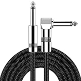
- [Low Noise High Fidelity] New bee guitar cord with 22AWG oxygen-free-copper center conductors provides enhanced signal transfer, the PVC insulation material and the high-density stranded copper double shield provide noise rejection for quiet operation
- [Ideal for Choice] New bee 1/4 straight and right angle metal connectors, ensures a secure connection in any instrument, pedal, or amp jack. L shape great fro on stage guitar stands
- [Wide Compatible] Suitable for mixer, power amplifier, microphone, effector, decoder, equalizer, electoronic organ, electric guitar, electric bass and other electronic equipments. Widely used in the installation of smart home, karaoke speaker, professional stage audio, etc
- [Easy Disassemble] Making incidental repairs quick and easy! Very nice, flexible cable
- [Dont Hesite to Order] 24 hours customer services and professional technology team are standing by
What does an fMRI brain map show?
An fMRI map usually shows locations where the measured MR signal changed in a way that matched a statistical model. Colors commonly represent the direction or strength of a contrast, such as greater signal during one condition than another, after correction choices and thresholds have been applied. The displayed voxels are therefore the end product of acquisition, preprocessing, registration, and statistical testing.
The signal is hemodynamic: it reflects blood-related changes that accompany neural activity rather than electrical firing itself. The relationship between neuronal events and hemodynamic responses varies, and the method has limits in both spatial and temporal resolution. A bright cluster can support a carefully qualified association between a region and a task under the study’s conditions; it cannot by itself identify the exact neurons involved, establish that the region is uniquely responsible, or show that it caused the behavior.
Resting-state maps use the same general type of MR time series but replace a task contrast with correlations in spontaneous fluctuations. They can reveal organized networks across distant regions, provided the analysis and preprocessing support that interpretation.
Recommended Free Tools
From scanner signal to a brain map: the data workflow
Informatics is part of the measurement story. The following stages explain why two analyses of apparently similar scans can produce different maps.
Rank #4

- Digital Family Calendar: BSIMB digital calendar wall touch screen puts an end to your chaotic life. Designed for busy families, the digital calendar keeps you organised and in step with your family. Assign different colours and tasks to each family member to make family scheduling easier and more intuitive.
- Easy Setup and Auto-Sync: Plug in, connect to WLAN and auto-syn your electric calendar in minutes. Digital wall calendar compatible with Google calendar, iCloud calendar, Outlook calendar, Cozi calendar and Yahoo calendar. Add events directly on the device or through the ecalendar app. Electronic calendar stays current whether you're at work or traveling—no manual updates needed.
- All-In-One Home Assistant: Smart calendar builds habits and encourage independence with personalised chores. Display daily meal plan to reduce the daily ‘What's for dinner?’ stress. To-do lists keep track of everything you want. Turning chores into valuable achievements with stars and rewards, making life easier and more enjoyable.
- Manage Anytime Anywhere: BSIMB electronic calendar for family stay connected with the powerful ecalendar app. Access and manage your digital wall calendar anytime, anywhere with free features like stars & rewards, photo screensaver, chore charts, meals plans, to-do lists etc. Electronic planner paid features like Magic Import tailor advanced tools for users with special needs.
- Stay in Touch with Loved Ones: Share photos or videos from your phone to electronic wall calendar with app, instantly update your life moment, and keep in touch with your family, especially those who are far away; you can also surprise your smart calendar digital wall by sending him a wish card; it's also great for keeping tech-illiterate elders in touch with the youngsters.
- Define the question and design the session. Researchers select participants, tasks or rest instructions, timing, behavioral responses, and the modality that can measure the target signal.
- Acquire raw measurements. The scanner or sensor records images, time series, electrical potentials, magnetic fields, or tracer emissions. Motion, physiological changes, scanner drift, and other noise are recorded along with the signal.
- Reconstruct the data. Computational reconstruction turns instrument measurements into three-dimensional images or time-resolved signals. For MRI, this may produce anatomical volumes and multiple functional or diffusion volumes.
- Correct and suppress noise. Analysts may correct for head motion, align volumes, account for timing differences, model physiological noise, and identify low-quality data. Each correction uses assumptions that can affect the final result.
- Register data to a reference. Images can be aligned to an individual’s anatomy or a common coordinate system so that regions can be compared across sessions and participants. Registration can introduce interpolation and alignment error.
- Estimate the scientific effect. Statistical models test task contrasts, relationships with behavior, group differences, or correlations between signals. Diffusion analyses fit models of water movement; tractography then propagates estimated orientations through neighboring voxels.
- Visualize and quantify. Results may be rendered as slices, cortical surfaces, network diagrams, tables of regional values, or time courses. A visualization is a summary of the analysis, not the raw measurement.
- Store, document, and share. Informatics systems support storage, querying, retrieval, comparison, and sharing. Formats such as NIfTI are commonly used for neuroimaging files, but software tools can differ in assumptions, defaults, and algorithms. Those differences create compatibility and reproducibility challenges unless processing steps and metadata are documented.
Because every stage can alter what is compared, a map should be read together with its acquisition parameters, preprocessing choices, statistical model, sample, and coordinate space.
Independent reader supportYour contribution helps us test, update, and keep practical guides available for everyone.What are brain maps used for?
Studying cognition and behavior
Researchers relate activity or connectivity measures to memory, language, attention, perception, movement, emotion, and decision-making. The strongest conclusions specify the task, comparison condition, population, and analysis rather than treating one activated region as a complete explanation of a mental process.
Characterizing healthy and disordered states
Imaging can reveal group-level anatomical, functional, or connectivity differences associated with neurological or psychiatric conditions. These findings help investigate mechanisms and potential biomarkers. A generic scan or online “brain map” is not, by itself, a diagnosis for an individual.
Tracking development and aging
Longitudinal and cross-sectional studies examine how anatomy, networks, and behavior vary across the lifespan. Interpretation must account for age, head motion, task performance, and other factors that change alongside brain organization.
Evaluating treatments and stimulation
Researchers use imaging to study pharmacological treatments and non-invasive stimulation methods, asking whether a measured signal or network changes after an intervention. An imaging change can document an association with treatment; it does not alone prove clinical benefit or explain the treatment’s mechanism.
Building connectivity resources
Large datasets allow researchers to estimate structural and functional connectivity across many participants, test individual differences, and develop standardized analyses. Shared data can support new questions without every team repeating the original scan session.
The Human Connectome Project as a large-scale example
The Human Connectome Project (HCP) illustrates how mapping combines modalities and participant information. Its goal was to acquire and share structural and functional connectivity data alongside behavioral measurements, demographic information, and participants’ DNA. This design supports analyses of how connectivity relates to behavior and how it differs between individuals and across the lifespan.
Do these 3 things before closing this tab:
1Repair Windows errors before they cause bigger problems2Fix the driver behind crashes, sound loss and screen glitches3Clear out junk files and repair common Windows errorsNIMH reports that the project mapped long-distance connections in 1,200 healthy young adults over five years — National Institute of Mental Health, 2022 (page last reviewed). The same account says the project began in 2010 after the National Institutes of Health awarded $40 million over five years — National Institutes of Health, 2010 (project award, as summarized by NIMH). These are historical project figures, not a current funding promise or a claim that the sample represents every population.
Standardized connectivity measures from projects such as HCP provide a foundation for disease-diagnosis research and other investigations. They do not turn a population map into an automatic clinical diagnosis, and they do not eliminate the need to validate findings in independent samples.
What brain maps cannot establish by themselves
- They are not complete pictures of the brain. Each modality samples particular signals at particular spatial and temporal scales. A map omits processes it was not designed to measure.
- fMRI is indirect. Its hemodynamic response is separated in time from the underlying neural events, and the relationship can vary across conditions and people.
- Resolution has trade-offs. A scan’s voxel size, sampling rate, acquisition sequence, and analysis determine which patterns can be distinguished. A smooth-looking image does not imply cellular resolution.
- Tractography is inferential. Crossing fibers, image noise, incomplete sampling, and modeling choices can produce false positives, false negatives, or uncertain endpoints. A drawn streamline is not a verified axon.
- Correlation is not causation. A region whose signal correlates with a task or behavior may participate in it, receive related input, or reflect another process. Causal claims require additional designs and evidence.
- Group averages can hide individual variation. A statistically reliable group map may not describe every participant, and a difference between groups may not classify a particular person accurately.
- Analysis choices matter. Motion handling, filtering, registration, region definitions, model specification, and statistical thresholds can change the apparent result. Independent replication and transparent methods are essential.
How to read a brain map responsibly
- Identify the modality and signal: anatomy, hemodynamic change, diffusion, electrical activity, magnetic field, tracer uptake, or a correlation.
- Check whether the image represents one participant, a group average, or a statistical contrast.
- Read the color bar, units, threshold, and coordinate system; color alone has no universal meaning.
- Ask what was measured directly and what was inferred by a model.
- Look for the task, comparison condition, sample characteristics, motion controls, and correction procedures.
- Separate an association from a causal or diagnostic claim.
- Check whether the result has been replicated or validated outside the original dataset.
Used this way, brain mapping is a powerful framework for connecting anatomy, activity, behavior, and estimated networks while keeping the evidence proportional to the measurement. Its maps are informative models of selected aspects of human brain organization—not literal, complete wiring diagrams of every neuron.
Quick Recap
Product prices and availability are accurate as of the date/time indicated and are subject to change. Any price and availability information displayed on Amazon at the time of purchase will apply.

