DNA can impose a helical, or chiral, organization on some of the water molecules next to it. A 2017 spectroscopy study detected that organization around two short DNA duplexes and called it a “chiral spine of hydration.” The result is evidence from an interface-sensitive measurement—not a photograph of a continuous water column, an observation inside living cells, or proof that the structure has a known biological function.
What is DNA’s chiral spine of hydration?
Chirality means that a structure has a handedness: its mirror image cannot be superimposed on it by ordinary rotation. The DNA double helix is chiral because its strands wind in one direction. Water molecules are individually achiral, but their hydrogen-bond network can become directionally organized when it is constrained by a chiral surface. In this case, the surface is the twisted chemical landscape of DNA.
As an Amazon Associate I earn from qualifying purchases.
The phrase “chiral spine of hydration” describes a population of water molecules near DNA whose collective vibrational response has the same handed character as the helix. It does not mean that every nearby molecule is locked into one permanent orientation. Water exchanges hydrogen-bond partners and reorients extremely rapidly. The claim is instead about a measurable, ensemble-averaged arrangement in the hydration shell: enough molecules adopt correlated orientations for a chiral optical signal to appear.
The Tool Desk
Outbyte PC Repair FREERepair Windows errors before they cause bigger problemsFix Now →Outbyte Driver Updater FREEFix the driver behind crashes, sound loss and screen glitchesFind Drivers →The 2017 work was led by Megan L. McDermott, Hannah Vanselous, Steven A. Corcelli and Poul B. Petersen. Their central conclusion was that DNA’s chirality is imprinted on nearby water. They also emphasized that the biological relevance of the observed arrangement was unknown.
#1 Best Overall

- Core Educational Tool: This DNA model kit features a realistic double helix structure that accurately represents the molecular structure of DNA, making it an effective biology DNA model for students to visually grasp genetic concepts and enhance their understanding of molecular biology
- Interactive DIY Assembly: Designed as a DIY double helix DNA model kit, it encourages hands-on learning and creativity by allowing students to assemble the model themselves, which promotes active engagement and better retention of scientific knowledge
- Collaborative Learning Enhancement: This molecular model kit is ideal for group projects and classroom demonstrations, enabling students to work together on building the DNA model, which fosters teamwork and collaborative understanding of genetic science
- Educational Science Awareness: the DNA double helix science kit serves to increase student interest in genetics and biology by providing a tangible, visual aid that deepens comprehension of complex biological structures and concepts
- Compact and Lightweight Design: Measuring 12.99 by 4.56 by 4.56 inches and weighing approximately 0.6 pounds, this gene model kit is portable and easy to handle, making it suitable for students and educators to carry and use in various learning environments
How the researchers detected the effect
The experiment used chiral sum-frequency generation (SFG) vibrational spectroscopy. SFG is a nonlinear optical method that combines two incident light fields at a surface or interface. When the frequencies are chosen appropriately, the emitted signal reports on molecular vibrations—in this study, the stretching vibration of the O–H bond in water.
Why the method can reveal orientation
An ordinary infrared spectrum mainly reports how strongly a bond absorbs light. SFG adds information about the direction in which molecular bonds are arranged at an interface. In a disordered, centrosymmetric bulk liquid, opposing molecular orientations tend to cancel in the SFG response. At an interface, or in an ordered layer, that cancellation is incomplete. Using polarization combinations that are sensitive to handedness lets researchers separate chiral contributions from the much larger achiral response.
The measured quantity is therefore an optical susceptibility associated with many water molecules, not a map of individual molecules. A peak or shoulder in the spectrum indicates that a group of O–H oscillators contributes coherently under the chosen measurement conditions. Interpreting that feature as a particular microscopic arrangement requires molecular and structural reasoning in addition to the raw spectrum.
What’s actually slowing this PC down?
Pick the symptom - the matching free tool is one click away.
What was placed in the measurement cell
The DNA was attached to a surface and immersed in aqueous solution. Each sample was a self-complementary, 24-base-pair duplex made from repeating base-pair units. The two constructs were:
Rank #2

- Educational Tool for Kids: This self-assembling dna display stand is an excellent educational tool for kids, helping them understand the structure of human genes in a fun and interactive way
- Interactive Learning Experience: The double helix model allows students to build their own dna structure, enhancing their understanding of molecular biology through hands-on activities
- High-Quality Material: Made from pp material, this dna model kit is designed to withstand repeated assembly and disassembly, making it a reliable teaching aid for classrooms and home use
- Perfect Size for Display: Measuring 4.56*4.56*12.99 inches, this dna model is the perfect size for classroom demonstrations and individual study, providing a clear and detailed representation of the dna structure
- Enhances Critical Thinking: This dna model kit not only teaches the basics of genetics but also helps develop critical thinking and problem-solving skills, learning more engaging and effective
| Sample | Sequence design | Conditions | Why it mattered |
|---|---|---|---|
| (GC)12 | Twelve alternating GC base pairs, forming a 24-base-pair duplex | Room temperature, 100 mM NaCl solution | Tests hydration around a GC-rich designed duplex |
| (AT)12 | Twelve alternating AT base pairs, forming a 24-base-pair duplex | Room temperature, 100 mM NaCl solution | Tests whether the response also appears for an AT-rich duplex and probes groove-dependent differences |
The authors described 100 mM sodium chloride as near-physiological ionic strength. That description is a useful qualification: the samples reproduced one important solution parameter, but they were still purified, surface-bound DNA constructs rather than chromosomes, cells or a complete cellular fluid.
What the spectra showed
A chiral response appeared for both DNA sequences
Both (GC)12 and (AT)12 produced a chiral water signal. This is important because it argues against the observation being peculiar to only one of the two tested base compositions. It does not establish that every DNA sequence, length, topology or chemical modification produces the same signal. The experiment sampled two carefully designed duplexes.
The chiral response was shifted to lower frequency
When the chiral spectrum was compared with the broader achiral hydration response, the chiral O–H feature was shifted toward lower vibrational frequencies. In hydrogen-bonded water, stronger hydrogen bonding generally weakens the effective O–H bond and moves its stretch to lower frequency. The researchers therefore interpreted the shift as evidence that the chiral population was, on average, more strongly hydrogen-bonded than the general hydration population.
Outdated Drivers Are Slowing You Down
One free scan finds every outdated or missing driver and matches the right update for your exact hardware.Free scan · exact hardware matchPC Slower Than It Used to Be?
A free scan shows the junk files, broken settings and background clutter dragging Windows down - then fixes them in one click.Free scan · Windows 10 & 11This is an interpretation of the vibrational response, not a direct measurement of a hydrogen-bond energy for each molecule. Frequency also depends on local geometry, coupling between oscillators, and the distribution of environments. The result supports a more strongly hydrogen-bonded subset of interfacial water, while the exact arrangement must be inferred.
Rank #3
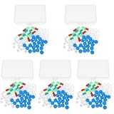
- Package includes five setsthe package list includes 5 x set of dna teaching model, providing multiple units for classroom rotation, group activities, or shared learning environments
- Package includes five setsthe package list includes 5 x set of dna teaching model, providing multiple units for classroom rotation, group activities, or shared learning environments
- Package includes five setsthe package list includes 5 x set of dna teaching model, providing multiple units for classroom rotation, group activities, or shared learning environments
- Package includes five setsthe package list includes 5 x set of dna teaching model, providing multiple units for classroom rotation, group activities, or shared learning environments
- Package includes five setsthe package list includes 5 x set of dna teaching model, providing multiple units for classroom rotation, group activities, or shared learning environments
An additional feature appeared in the AT sample
The AT duplex showed a feature the authors attributed to non-hydrogen-bonded O–H groups. They suggested that tighter confinement in the AT minor groove could help explain why some O–H bonds appeared less involved in the usual hydrogen-bond network. This proposal links the spectrum to the different width and chemical environment of DNA grooves, but it remains a molecular explanation for the observed feature rather than an image of water inside the groove.
Where the water is likely to be located
DNA presents two grooves with different widths and chemical patterns. The minor groove is narrower and can create a more confining environment for solvent. The authors concluded that the detected chiral response was likely dominated by water near the minor groove. They could not completely exclude a contribution from water near the major groove, because the optical experiment measures the interfacial response of the sample rather than assigning every signal to one groove with absolute certainty.
A useful mental model is a hydration shell whose molecules have different degrees of freedom. Some water remains relatively bulk-like; some is constrained by phosphate groups, base edges and groove geometry; and a subset may form correlated, handed hydrogen-bond arrangements. The spectroscopy highlights the subset that contributes to the chiral response. It does not imply that the entire surrounding solution has become a rigid helix.
What “templated by DNA” means
DNA does not need to make covalent bonds to every water molecule to template an arrangement. Electrostatic fields, hydrogen-bond donors and acceptors, steric confinement and the repeating geometry of the helix can bias how water molecules orient and connect. Because those influences repeat along a chiral scaffold, the average orientation of nearby water can acquire a handed component.
Rank #4

- √Principle: In a double-stranded DNA molecule, A=T, G=C. That is: A + G = T + C or A + C = T + G;
- √Interlocking pieces connect to form the double helix shape and show how molecules split at the center of the base pairs
- √Completed model measures 33cm [13"] high
- √Make learning come alive and build creativity with this hands-on and interactive science kit!
- √Note: Recommended for ages 14+
Water remains dynamic under these conditions. Molecules can exchange between hydration layers and bulk solution, and the experiment averages over that motion during the measurement. “Templated” therefore means that DNA’s structure biases the statistical distribution of orientations and hydrogen-bonding environments strongly enough to generate a chiral signal, not that a motionless molecular chain has been observed.
What the study established—and what it did not
| Supported by the experiment | Not established by the experiment |
|---|---|
| A chiral O–H vibrational response from water was detected at an interface containing DNA. | A continuous, visible water column running along the helix was not imaged. |
| The response occurred for both surface-bound 24-base-pair duplexes, (GC)12 and (AT)12. | All genomic sequences, DNA lengths, supercoiled forms or chemically modified DNAs were not tested. |
| The chiral response was at lower frequency than the general achiral hydration response, consistent with stronger hydrogen bonding in that population. | The spectra do not by themselves prove the exact orientation of every water molecule or provide a unique three-dimensional structure. |
| The signal was likely dominated by minor-groove hydration, with major-groove water not entirely excluded. | The measurement was not performed inside living cells or in a complete cellular environment. |
| The method can probe DNA hydration without fluorescent or chemical labels. | A biological function, disease mechanism, or demonstrated change in drug binding was not shown. |
Why the result matters for DNA chemistry
Hydration is part of DNA structure rather than a passive coating. Water and ions help screen the negatively charged phosphate backbone, stabilize grooves and influence the flexibility of the helix. Many proteins, small molecules and drugs encounter DNA through a combination of direct contacts and the displacement or rearrangement of water. A way to measure the handed component of hydration could therefore add information that conventional bulk spectroscopy misses.
The value of the work is methodological as much as structural. Chiral SFG can examine an interfacial hydration environment without attaching a fluorescent tag that might perturb the sample. Because the method is sensitive to orientation and symmetry, it can distinguish a chiral contribution even when the total amount of water near DNA is much larger than the specially organized fraction.
Quick wins for a faster PC:
Repair Windows errors before they cause bigger problemsFix Now →Fix the driver behind crashes, sound loss and screen glitchesFind Drivers →That potential does not turn the 2017 observation into a demonstrated biological mechanism. The authors explicitly identified biological relevance as an open question. A correlation between a chiral hydration signal and a cellular outcome would require separate experiments under conditions where both the hydration state and the biological process are measured.
Best Value

- Intuitive teaching tools to improve learning effects: This DNA double helix structure model is designed for middle school biology and high school courses, and can intuitively display the complexity of genes and molecular structures. Through assembly of the model, students can have a deeper understanding of the basic structure of DNA and its role in the transmission of information, and enhance classroom interactivity and participation
- High-precision restoration, realistic details: The model is made of plastic materials, and each component is carefully designed to accurately simulate the molecular structure, helping students to quickly identify each part and establish a clear visual memory
- Flexible combination, cultivate hands-on ability: Provide a variety of detachable and recombinable components to encourage students to build the DNA double helix structure by themselves. This process not only deepens the understanding of knowledge points, but also effectively exercises students spatial thinking ability and hands-on practical skills, which is classroom teaching demonstrations and research projects
- Safe and reliable: The sturdy and design allows the model to be reused between multiple semesters, reducing resource waste, and is also convenient for school or family preservation and management. It is an ideal educational investment, both practical and educational
- DNA double helix structure model kit, it is made of plastic material, reliable and safe, easy to assemble and disassemble. Professional DNA double helix structure model makes your easy understanding of terminology, it is a nice science educational teaching instrument toy
Questions future experiments would need to answer
Does sequence control the signal?
Testing only GC- and AT-repeating duplexes cannot determine how mixed sequences, unusual base pairs, methylation, mismatches or damage affect the chiral response. A systematic sequence series would be needed, with the DNA length, surface coverage, salt concentration and temperature controlled so that spectral differences could be assigned to sequence rather than to sample preparation.
How do ions and other solutes change hydration?
Sodium chloride was held at 100 mM in the reported measurements. Other ions can bind differently to phosphates and grooves, and multivalent ions can alter DNA conformation. Comparing salt identities and concentrations could show whether the chiral water population is robust or highly sensitive to electrostatic screening.
What happens when proteins or drugs bind?
A ligand entering a groove may displace water, reorganize hydrogen bonds or change the DNA conformation. Chiral SFG could be used to follow those changes at a model interface. Such an experiment would still need controls and independent binding measurements before a spectral change could be identified as the cause of a biological effect.
Recommended Free Tools
Does the arrangement persist in crowded environments?
Cells contain proteins, metabolites, nucleic acids and variable concentrations of ions. Surface-bound duplexes in a simple solution isolate the DNA–water interaction, which is useful for interpretation but does not reproduce molecular crowding. Measurements with controlled crowding agents or reconstituted complexes would test how transferable the observation is.
How to interpret headlines about a “water spine”
- Read “spine” as a collective pattern. The term refers to an averaged chiral hydration response, not a solid rod of water.
- Check the measurement. The evidence came from chiral SFG vibrational spectroscopy of O–H stretches at a DNA–water interface.
- Check the sample. The reported constructs were surface-bound, self-complementary 24-base-pair duplexes in room-temperature 100 mM NaCl solution.
- Separate observation from interpretation. A lower-frequency chiral feature supports stronger hydrogen bonding in that population; groove localization and detailed molecular orientation are inferred.
- Do not add a biological claim. The study did not establish a function in cells, a universal property of DNA or a proven effect on drug action.
Publication context
The paper was received on March 6, 2017, published online on May 24, 2017, and appeared in the July 26, 2017 issue of ACS Central Science. Its abstract summarizes the result as evidence that DNA’s chirality is reflected in nearby water while noting that the biological relevance of a chiral spine of hydration remains unknown. That qualification is central, not a minor footnote.
Bottom line
The 2017 study provides a careful piece of physical chemistry: two designed DNA duplexes produced a chiral water vibrational response under controlled solution conditions, and that response was consistent with a more strongly hydrogen-bonded, DNA-templated hydration population probably concentrated near the minor groove. It does not show a literal water helix in living cells or reveal what the structure does biologically. The most defensible description is a spectroscopically detected chiral organization of interfacial water whose function remains an open research question.
Quick Recap
Product prices and availability are accurate as of the date/time indicated and are subject to change. Any price and availability information displayed on Amazon at the time of purchase will apply.

