Do these 3 things before closing this tab:
1Clear out junk files and repair common Windows errors2Scan for outdated or missing drivers - takes under a minute3Repair Windows errors before they cause bigger problemsNeither architecture is universally better. Single-objective light-sheet microscopy places one sample-proximal objective near the specimen for both illumination and fluorescence collection, which can simplify mounting and improve physical access. Conventional light-sheet microscopy generally uses separate, often perpendicular, objectives: one creates the light sheet and the other detects fluorescence. That separation gives independent control of the illumination and detection paths, but it can restrict holders, chambers and specimen geometry.
Choose by the experiment rather than by the label. Compare the actual system’s usable depth, numerical aperture, resolution, sensitivity, field of view, scan range, volume rate, phototoxicity, multi-view capability and alignment workflow against your specimen and acquisition plan. The sections below explain what each architecture changes, where the trade-offs arise and how to evaluate two specific instruments without treating one instrument’s specifications as a universal standard.

As an Amazon Associate I earn from qualifying purchases.
What the two architectures mean
Conventional light-sheet: separate illumination and detection objectives
In the conventional arrangement, an illumination objective forms a thin sheet of excitation light through the specimen. A second objective, commonly positioned at approximately 90 degrees to the first, collects the fluorescence from the illuminated plane and images it onto a camera. The orthogonal geometry is often called dual-objective light-sheet microscopy.
Because the paths are separate, a designer can optimize the illumination and collection optics independently. The detection objective can be selected for collection numerical aperture, correction and working distance without also having to create the sheet. Illumination shaping, scanning and filtering can likewise be engineered on their own optical path. The cost is physical: the sample must be accessible from both directions, and the chamber, holder or mounting method has to leave those paths clear.
#1 Best Overall

- [Ultra-Lightweight Ear Buds Designed for Small Ears] Each earbud weighs only 3.7g and features a compact, ergonomic in-ear design made especially for small ears. Secure, low-profile, and comfortable for workouts, all-day wear or overnight sleeping.The ultra-slim profile sits flush in your ear with no hard edges poking or rubbing, so you can even sleep on your side without discomfort.
- [Immersive Stereo Sound with TOZO OrigX Technology] TOZO OrigX tuning delivers clear vocals, balanced mids, and natural stereo sound for music, podcasts, and videos.
- [Long Battery Life for Daily Use] Get up to 7 hours of playtime on a single charge, with up to 32 hours total using the charging case—ideal for workdays, commuting, and extended listening sessions.
- [Bluetooth 5.3 & Stable Connection] Bluetooth 5.3 provides fast pairing, stable wireless performance, and reduced dropouts as you move around home or office.
- [Deep Bass with Clear Vocals] High-performance drivers produce punchy bass while keeping vocals clean and detailed for everyday listening.
Single-objective light-sheet: one objective close to the sample
A single-objective instrument uses one objective near the specimen for both delivering the sheet and collecting fluorescence. Oblique-plane microscopy (OPM) is a prominent family of this design. The objective views an oblique plane, while remote objectives and relay optics transfer the emitted image to a camera.
Having only one primary objective beside the sample can make planar or otherwise familiar mounting arrangements easier. It does not mean that the entire microscope is optically simple. Remote focusing, tertiary or relay objectives, image rotation and deskewing can move complexity away from the specimen and into the optical train and software.
Why “single” does not automatically mean simpler or better
Single-objective systems cannot place the sheet at a right angle to the primary objective’s own axis. The oblique image plane therefore cannot usually be recorded directly as an undistorted, in-focus rectangle on a flat detector. Relaying, geometric transformation and, in many implementations, computational correction are required. Earlier OPM implementations could sacrifice primary-objective detection numerical aperture, resolution, sensitivity or field of view; custom remote-focus designs have reduced some of those penalties without eliminating every engineering trade-off.
Conversely, “conventional” does not guarantee a particular resolution, depth or speed. Objective choices, refractive-index matching, sheet thickness, camera format, scanning hardware and sample preparation determine the practical result.
How the optical paths differ in practice
Dual-objective path
- An illumination source and shaping optics prepare the excitation beam.
- The illumination objective focuses that beam into a sheet through the specimen.
- The detection objective, viewing from another direction, collects fluorescence from the sheet.
- Emission filters and a camera record the selected plane while the sheet or specimen is scanned to build a volume.
The independent paths make it straightforward to change illumination and detection components separately. They also require stable alignment between two objectives and enough open space around the sample for both.
Single-objective OPM path
- The sample-proximal objective delivers illumination at an oblique angle and collects fluorescence.
- Remote objective(s) and relay optics refocus or reimage the oblique plane.
- The camera records the relayed image, often with rotation or deskewing needed to represent the specimen in conventional coordinates.
- Scanning may move the stage, the beam, a galvo mirror or several of these together.
This arrangement can leave the top or sides of a mounted sample more accessible. However, every relay contributes aberration, alignment sensitivity and potentially a restriction on field or numerical aperture.
Rank #2

- 【IP68 Waterproof Multifunctinal Smart Watch】Torjalph fitness activity tracker smart watch is IP68 waterproof technology ,you can wear it when swimming,handwashing,it's rain-proof which means that you perform multiple exercise modes outside without worrying about sweat or rain corroding your fitness smart watch,and this watch equipped with a 230 mAh large capacity battery magnetic charging ,and this sports activity tracker watch can be used 5-7 days If fully charged.
- 【Larger 1.83'' Full Screen Watch & Multi-dial Selection】1.83 inch full HD color touch screen smart watch for men / women provides excellent 240*284 resolution image quality ,This Bluetooth smart watch offers a better touch experience and remarkable immersion and ,more than 200 watch faces for you to choose from App"Da Fit ", you can change the dial style according to your mood to wear keeping the world on your wrist
- 【Accurate Activity Tracker Smart Watch 】sports watch activity tracker uses sweat and dust resistant breathable silicone strap, our Fitness Tracker Smart Watch for Android iOS Phones supports 12 sports modes,walking,running,bicycle,swimming,badminton,basketball,football,rope skipping. This bluetooth calls smart watch for men women is lightweight and stylish, bringing more convenient and relaxing sports experience while also providing more protection.
- 【Your 24 Hour Personal Daily Health Monitor】Fitness watch for women men full touch screen IP67 waterproof bluetooth smartwatch for Android iOS Phones with heart rate blood pressure monitor multifunction Andriod smart watch with steps counter sleep monitor sports fitness watch, monitoring blood oxygen saturation allows you to understand your physical condition in real time to track fitness progress ,to know your health status in time.
- 【More Functions Bluetooth Call Smart Watch】This fitness activity smart watch full screen for iPhone and Android phones includes a bunch of new features including:The bluetooth smart watch can hang up the phone, receive and display WhatsAPP /Text /Facebook /Twitter /Messenger reminder.Support sedentary reminder, alarm clock, stopwatch, timer,weather information,music control, remote camera control compatible with Android 5.0 and above or iOS 9.0 and above smartphones.
Head-to-head comparison
| Decision axis | Single-objective architecture | Conventional dual-objective architecture |
|---|---|---|
| Objectives beside the specimen | One primary objective performs illumination and collection; remote objectives may relay the image. | Separate objectives form the sheet and collect fluorescence. |
| Sample access | Often more compatible with planar or familiar mounting because fewer objectives surround the specimen. | Requires optical access from two directions; chambers and holders must keep both paths clear. |
| Optical independence | Illumination and detection share the primary objective, so some parameters are coupled. | Illumination and detection paths can be optimized independently. |
| Image geometry | Oblique-plane imaging normally needs relay optics and geometric or computational correction. | Orthogonal geometry is naturally aligned with a conventional camera plane. |
| Working distance and usable depth | Set by the primary objective, relay design, sample interfaces and aberration correction. | Set independently by the two objectives, but both must reach the intended plane through the mounting arrangement. |
| Resolution and sensitivity | Implementation-dependent; relay and primary-objective constraints can reduce performance, while custom designs can recover some capability. | Independent detection-path optimization can be advantageous, subject to objective and sample limits. |
| Field of view | Can be limited by the primary objective or relay; custom systems may provide large fields. | Determined mainly by detection objective, camera and sheet uniformity over the specimen. |
| Scan range and speed | Depends on stage, galvo and relay geometry; oblique-plane correction may add processing overhead. | Depends on sheet scanning, stage travel, camera and synchronization of the two paths. |
| Multi-view coverage | Not inherent; available only if the instrument is designed to illuminate or view from additional directions. | Also not inherent; multiple views require additional optical paths or a sample-rotation strategy. |
| Alignment and maintenance | Fewer sample-side objectives but potentially demanding remote-focus and relay alignment. | More objectives around the sample, with alignment of two primary paths. |
The table describes architectural tendencies, not guaranteed specifications. A real comparison should use measurements made with the same specimen, fluorophores and acquisition task.
Sample access, mounting and usable depth
When access is the deciding constraint
A dual-objective system needs a clear route to the sample from the illumination and detection directions. That can be practical for a cleared organ or a purpose-built chamber, but awkward for a specimen on a standard slide, a culture device with a solid base or a preparation that must remain open from above. The objectives, immersion medium and holder all have to coexist without clipping the sheet or blocking the collection cone.
A single objective can approach the specimen from one side, which is useful when the sample is mounted in a plane or when the opposite side must remain available for manipulation. This is an access advantage, not proof that every sample will image more deeply. The objective still has a finite working distance, and coverslips, gels, walls and refractive-index transitions can consume that distance or introduce aberration.
Open-top and mismatched interfaces
Open-top dual-objective designs can improve physical access for some preparations, but modeled ODO/NOSO open-top comparisons show that the benefit can come with reduced usable axial range and more demanding high-NA correction across mismatched optical interfaces. Treat those results as modeled design analysis rather than a universal experimental head-to-head. Ask a facility for the usable, corrected depth in the exact chamber and medium you will use, not only the objective’s nominal working distance.
Questions to ask about depth
- What is the objective’s working distance after the coverslip, chamber wall or mounting gel is included?
- Over what axial range are resolution and sensitivity maintained, rather than merely detectable?
- What refractive index does the objective correction assume, and how is mismatch handled?
- Does the reported depth apply to the center of a cleared sample, a near-surface specimen or a particular immersion medium?
Resolution, sensitivity and field of view
Numerical aperture is necessary but not sufficient
Higher collection numerical aperture generally supports finer detail and more efficient fluorescence collection, but the final result also depends on wavelength, aberrations, sheet thickness, camera sampling, signal background and the depth at which the measurement is made. In a single-objective design, the primary objective must support both illumination and detection, and relays must preserve the image. In a dual-objective design, the detection path can be optimized separately, which may simplify high-NA collection.
Windows Errors? Fix Them Before They Spread
Repair common Windows errors and clear accumulated junk for a smoother, more stable PC - no reinstall needed.Free scan · no reinstallCrashes, No Sound, or Screen Glitches?
Random freezes, missing sound and display glitches usually trace back to one bad driver. Find and replace yours safely.Free scan · under a minuteDo not compare a headline lateral-resolution number from one instrument with an axial number from another. Require the point-spread function (PSF), the bead size and refractive-index conditions, the depth in the sample and the analysis method. Ask whether the value is measured, simulated or inferred from optical specifications.
Rank #3
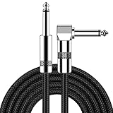
- [Low Noise High Fidelity] New bee guitar cord with 22AWG oxygen-free-copper center conductors provides enhanced signal transfer, the PVC insulation material and the high-density stranded copper double shield provide noise rejection for quiet operation
- [Ideal for Choice] New bee 1/4 straight and right angle metal connectors, ensures a secure connection in any instrument, pedal, or amp jack. L shape great fro on stage guitar stands
- [Wide Compatible] Suitable for mixer, power amplifier, microphone, effector, decoder, equalizer, electoronic organ, electric guitar, electric bass and other electronic equipments. Widely used in the installation of smart home, karaoke speaker, professional stage audio, etc
- [Easy Disassemble] Making incidental repairs quick and easy! Very nice, flexible cable
- [Dont Hesite to Order] 24 hours customer services and professional technology team are standing by
DaXi: a design-specific single-objective example
The 2022 DaXi paper illustrates what a custom OPM instrument can achieve, but its values are not specifications for single-objective microscopy as a category. The system used a 20× primary objective and a custom tertiary objective with NA 1.0. The authors report a field of view of up to 750 micrometres.
| DaXi reported item | Value and qualification |
|---|---|
| Primary objective | 20×, in the authors’ instrument |
| Custom tertiary objective | NA 1.0 |
| Maximum reported field of view | Up to 750 µm |
| Stage travel in the reported configuration | Up to 75 mm |
| Galvo-only scanning range | About 300 µm |
| Measured bead PSF, principal axis 1 | 479.9 ± 28.0 nm |
| Measured bead PSF, principal axis 2 | 379.2 ± 20.9 nm |
| Measured bead PSF, principal axis 3 | 1,864.9 ± 174.3 nm |
Those PSF values were measured under the authors’ setup and analysis. They should be used to understand the instrument’s demonstrated behavior, not to predict the resolution of another OPM system or to rank the two architectures in general.
Field of view and stitching
A large field can reduce tiling, but only if illumination is sufficiently uniform and resolution remains acceptable across that field. When a specimen exceeds one view, compare not just the nominal field size but tile overlap, stitching time, registration accuracy and the loss of temporal resolution while the system moves between tiles.
Quick wins for a faster PC:
Scan for outdated or missing drivers - takes under a minuteDriver Scan →Repair Windows errors before they cause bigger problemsFix Now →Scan range, acquisition speed and volume size
Light-sheet imaging can scan a plane with a moving sheet, a moving specimen, beam steering or a combination. The useful metric is the completed volume at the required signal-to-noise ratio, not the fastest camera frame rate in isolation.
DaXi combined continuous stage motion with compensating galvo motion. In that configuration, the stage could travel up to 75 mm, while galvo scanning alone covered about 300 µm. The two ranges serve different purposes: a galvo can provide rapid fine scanning over a limited distance, whereas stage travel supports larger volumes at a different mechanical and synchronization cost.
For any candidate instrument, document:
- Maximum continuous scan distance and the range over which focus and illumination remain valid.
- Plane rate, frame rate and the exposure time used to reach the required signal-to-noise ratio.
- Time to acquire a complete volume, including acceleration, settling, tile changes and channel switches.
- Whether deskewing, deconvolution or multi-view registration can run during acquisition or must be performed afterward.
Multi-view imaging and occlusion
Scattering, absorption and refraction can hide structures behind other structures. Viewing or illuminating from another direction can reveal information that one direction misses, but multi-view capability is an instrument feature rather than a consequence of using one objective.
Rank #4

- Digital Family Calendar: BSIMB digital calendar wall touch screen puts an end to your chaotic life. Designed for busy families, the digital calendar keeps you organised and in step with your family. Assign different colours and tasks to each family member to make family scheduling easier and more intuitive.
- Easy Setup and Auto-Sync: Plug in, connect to WLAN and auto-syn your electric calendar in minutes. Digital wall calendar compatible with Google calendar, iCloud calendar, Outlook calendar, Cozi calendar and Yahoo calendar. Add events directly on the device or through the ecalendar app. Electronic calendar stays current whether you're at work or traveling—no manual updates needed.
- All-In-One Home Assistant: Smart calendar builds habits and encourage independence with personalised chores. Display daily meal plan to reduce the daily ‘What's for dinner?’ stress. To-do lists keep track of everything you want. Turning chores into valuable achievements with stars and rewards, making life easier and more enjoyable.
- Manage Anytime Anywhere: BSIMB electronic calendar for family stay connected with the powerful ecalendar app. Access and manage your digital wall calendar anytime, anywhere with free features like stars & rewards, photo screensaver, chore charts, meals plans, to-do lists etc. Electronic planner paid features like Magic Import tailor advanced tools for users with special needs.
- Stay in Touch with Loved Ones: Share photos or videos from your phone to electronic wall calendar with app, instantly update your life moment, and keep in touch with your family, especially those who are far away; you can also surprise your smart calendar digital wall by sending him a wish card; it's also great for keeping tech-illiterate elders in touch with the youngsters.
The DaXi authors alternated illumination and viewing directions to obtain complementary views and reduce occlusion effects. A different single-objective system may provide only one view. A dual-objective system may also support multiple views if it includes additional paths, sample rotation or a suitable scanning design. Confirm the actual mechanism, angular range, registration method and acquisition-time penalty.
Free tools Windows power users keep installed
One-click scans. No signup required.
Optical and computational complexity
Where dual-objective systems are demanding
- Two objectives must remain aligned with the same illuminated plane.
- The sample chamber must preserve access and refractive-index conditions for both paths.
- Changing the specimen or holder can require re-alignment of illumination and detection.
- Large objectives and immersion hardware can limit working space around the sample.
Where single-objective systems are demanding
- Remote objectives and relays must preserve focus, magnification and numerical aperture.
- Oblique images require geometric correction, deskewing or both before quantitative analysis.
- Relay aberrations and vignetting can vary with depth and field position.
- Primary-objective compromises may couple illumination quality to detection performance.
Count the full workflow: alignment time, calibration beads, refractive-index matching, correction files, storage and analysis. A system with fewer sample-side parts may still require more specialized alignment or software expertise.
Phototoxicity and bleaching
Plane illumination confines excitation to a sheet, which can reduce out-of-focus exposure compared with wide-field illumination. That is a general light-sheet advantage, not a guaranteed advantage of single-objective over dual-objective geometry. Phototoxicity and bleaching depend on illumination dose, sheet thickness, dwell time, fluorophore, wavelength, scan strategy and the temporal resolution required.
Compare the dose needed for the same biological endpoint: the same volume, signal-to-noise ratio, frame interval and number of channels. A faster scan that produces insufficient signal may force higher illumination power or repeated acquisitions, erasing an apparent speed advantage.
How to choose between two actual instruments
- Define the specimen geometry. Record dimensions, mounting medium, coverslip or chamber walls, access needed for manipulation and whether the preparation can expose two sides.
- Set the biological requirement. Specify the smallest structure, depth, field, volume, time interval and number of channels that must be captured.
- Request depth-qualified performance. Ask for measured lateral and axial PSFs, collection NA, sensitivity and usable depth in a comparable sample or test target.
- Calculate the acquisition burden. Include scan travel, camera exposure, settling, tile overlap, channel changes, multi-view passes and processing time.
- Check optical interfaces. Verify refractive-index assumptions, immersion medium, correction collars, chamber materials and whether the reported performance changes with depth.
- Evaluate views and occlusion. Determine whether one direction is sufficient or whether alternating views, rotation or additional objectives are required.
- Audit the workflow. Include alignment, calibration, deskewing, deconvolution, registration, data storage and staff training.
- Run a specimen-specific pilot. Use representative size, labeling density and optical properties. Measure the endpoint that matters instead of extrapolating from a brochure specification.
Which architecture fits common experimental situations?
Planar samples and restricted underside access
A single-objective arrangement is often worth considering when the sample must remain on a familiar plane or when a second objective cannot approach the opposite side. Verify that the primary objective’s working distance and the relay’s field cover the full region of interest.
The Tool Desk
Outbyte PC Repair FREEClear out junk files and repair common Windows errorsFree Scan →Outbyte Driver Updater FREEFix the driver behind crashes, sound loss and screen glitchesFind Drivers →Samples that permit dedicated two-sided mounting
A conventional dual-objective system can be attractive when a purpose-built chamber provides clear access and independent optimization of sheet formation and fluorescence collection is important. Confirm that the holder does not clip either numerical-aperture cone.
Large, scattering specimens
Prioritize usable depth, multi-view coverage and registration over the architecture label. A single-objective system with only one direction may leave occluded structures unresolved; a dual-objective system without additional views has the same limitation.
High-speed volumetric imaging
Compare completed-volume time and signal at the required depth. Stage-plus-galvo schemes, camera exposure, synchronization and processing can dominate the result in either architecture.
Common comparison mistakes
- Assuming fewer objectives means less complexity. Remote relays and oblique-plane correction can be substantial.
- Using nominal working distance as usable depth. Interfaces and aberration correction may reduce the range with acceptable performance.
- Comparing unmatched PSFs. Depth, bead size, wavelength and analysis method must be aligned.
- Treating one custom instrument as representative. DaXi’s 20× objective, NA 1.0 tertiary objective, 750-µm field and reported PSF belong to that configuration.
- Calling multi-view inherent to single-objective designs. It requires additional optical or mechanical capability.
- Counting camera frames instead of useful volumes. Include exposure, travel, settling, tiles, channels and processing.
- Ignoring sample handling. A theoretically strong optical path is not useful if the specimen cannot be mounted, changed or kept viable.
- Assuming light-sheet phototoxicity is architecture-specific. Dose is determined by the full illumination and acquisition protocol.
A practical specification checklist
Before selecting a system or planning a facility evaluation, request a written specification sheet covering:
Recommended Free Tools
- Primary and remote objective magnification, NA, working distance and immersion medium.
- Sample chamber dimensions, access directions and supported mounting materials.
- Measured lateral and axial PSFs, with depth and test conditions.
- Usable field of view, uniformity and any tile-stitching requirement.
- Illumination sheet thickness, power range and scan method.
- Stage and galvo travel, acceleration, synchronization and repeatability.
- Frame rate, volume rate and signal-to-noise at the intended depth.
- Available viewing directions, multi-view registration and occlusion handling.
- Deskewing, deconvolution and analysis software requirements.
- Calibration procedure, alignment time and maintenance of relay or immersion optics.
Bottom line
Choose single-objective light-sheet microscopy when sample access, planar mounting or a standard footprint is the dominant constraint and your lab can support oblique-plane relays and correction. Choose conventional dual-objective geometry when the specimen can be mounted with two-sided optical access and independent illumination and detection optimization is more valuable than that access.
For a defensible decision, compare the two complete instruments on the same specimen: corrected depth, resolution, sensitivity, field, volume rate, scan range, dose, views and workflow. Architecture establishes the trade-offs; the implementation determines the result.
Quick Recap
Product prices and availability are accurate as of the date/time indicated and are subject to change. Any price and availability information displayed on Amazon at the time of purchase will apply.

