Outdated Drivers Are Slowing You Down
One free scan finds every outdated or missing driver and matches the right update for your exact hardware.Free scan · exact hardware matchPC Slower Than It Used to Be?
A free scan shows the junk files, broken settings and background clutter dragging Windows down - then fixes them in one click.Free scan · Windows 10 & 11Single-cell RNA sequencing (scRNA-seq) tells you which cell types and transcriptional states are present; spatial transcriptomics tells you where those states occur in the embryo. scRNA-seq usually requires dissociation into cells or nuclei, so native positions are lost. Spatial assays preserve a coordinate in tissue, but their measurements may represent a mixed spot or, in imaging systems, an individual cell or subcellular region.
Neither method is universally better. Choose according to the biological question, required resolution, tissue and sectioning constraints, field of view, throughput, and computational workload. For many developmental studies, scRNA-seq discovers and annotates cell states while spatial data maps those states back onto embryonic anatomy.

As an Amazon Associate I earn from qualifying purchases.
What is the difference between scRNA-seq and spatial transcriptomics?
Both methods measure RNA expression, but they preserve different information. scRNA-seq measures a transcriptome for each captured cell or nucleus after tissue has been dissociated. The output is a cell-by-gene matrix that is well suited to finding populations, developmental states, trajectories, and gene-regulatory programs. The trade-off is that the dissociation process normally removes the coordinates that tell you where each cell sat in the embryo.
Spatial transcriptomics measures RNA while retaining a relationship to a position in a tissue section. A coordinate can correspond to a barcode-containing spot that captures RNA from several cells, or to a cell-sized or subcellular signal detected by imaging. Spatial data therefore answer questions such as whether a state is confined to a germ layer, boundary, organ primordium, or signaling center.
#1 Best Overall

- [Ultra-Lightweight Ear Buds Designed for Small Ears] Each earbud weighs only 3.7g and features a compact, ergonomic in-ear design made especially for small ears. Secure, low-profile, and comfortable for workouts, all-day wear or overnight sleeping.The ultra-slim profile sits flush in your ear with no hard edges poking or rubbing, so you can even sleep on your side without discomfort.
- [Immersive Stereo Sound with TOZO OrigX Technology] TOZO OrigX tuning delivers clear vocals, balanced mids, and natural stereo sound for music, podcasts, and videos.
- [Long Battery Life for Daily Use] Get up to 7 hours of playtime on a single charge, with up to 32 hours total using the charging case—ideal for workdays, commuting, and extended listening sessions.
- [Bluetooth 5.3 & Stable Connection] Bluetooth 5.3 provides fast pairing, stable wireless performance, and reduced dropouts as you move around home or office.
- [Deep Bass with Clear Vocals] High-performance drivers produce punchy bass while keeping vocals clean and detailed for everyday listening.
| Decision point | scRNA-seq | Spatial transcriptomics |
|---|---|---|
| Primary output | Expression profile for each dissociated cell or nucleus | Expression associated with a coordinate in preserved tissue |
| Native position | Generally lost during dissociation | Retained, with accuracy determined by assay and tissue handling |
| Resolution | Usually cell-level for captured cells | Method-dependent: mixed spots, cell-level signals, or subcellular imaging |
| Main strength | Discovering cell types, subpopulations, and transcriptional programs at scale | Mapping expression patterns and cell states onto anatomy |
| Main limitation | Cannot by itself show where a cell came from in the embryo | Resolution, transcript coverage, field of view, and imaging or diffusion effects vary by platform |
| Typical interpretation | “What states and programs exist?” | “Where are those states and programs located?” |
The distinction is not absolute. Some spatially resolved methods produce near-single-cell data, while some spot-based assays contain RNA from portions of several neighboring cells. Treat “spatial transcriptomics” as a family of measurement models rather than one fixed technology.
Why the choice matters especially in embryos
Embryos change rapidly in both cell identity and geometry. A transient transcriptional state may appear only at a particular stage, and its position relative to a signaling source can determine its fate. Dissociation can reveal that the state exists but cannot establish whether it formed at a tissue boundary, migrated there, or was distributed throughout the embryo. Conversely, a spatial map can show a pattern without providing enough transcriptomic depth to separate closely related states.
Embryo samples also impose practical constraints. The specimen may be small, limited in number, and difficult to orient consistently. Sectioning can divide a three-dimensional structure into partial views, and RNA quality can change during fixation, permeabilization, and imaging. A design that works for a large adult tissue may not provide adequate field of view or molecular sensitivity for an early embryo.
Do these 3 things before closing this tab:
1Fix the driver behind crashes, sound loss and screen glitches2Clear out junk files and repair common Windows errors3Scan for outdated or missing drivers - takes under a minuteHow each method works
scRNA-seq workflow
- Collect and stage the embryos. Record species, developmental stage, genotype, treatment, and dissection orientation. Developmental timing is a biological variable, not just a metadata field.
- Dissociate tissue or isolate nuclei. Enzymatic and mechanical steps release cells; nuclei preparation can be preferable when cells are fragile or tissues are difficult to dissociate. Either approach can introduce selective loss or stress responses.
- Capture individual cells or nuclei with barcodes. Each partition receives a molecular barcode, allowing transcripts from that partition to be assigned to one cell or nucleus.
- Sequence and build a count matrix. Reads are aligned or pseudo-aligned to a reference, then summarized as counts by gene and barcode.
- Filter and analyze. Researchers remove low-quality barcodes, identify doublets, normalize counts, reduce dimensions, cluster cells, and annotate populations using marker genes and developmental knowledge.
The resulting matrix supports comparisons across thousands of cells and can expose rare populations that would be difficult to isolate manually. However, the matrix does not retain the original coordinates unless the experiment deliberately records a coarse anatomical subdivision before dissociation.
Spatial transcriptomics workflow
- Preserve and orient the tissue. Fixation, freezing, embedding, and sectioning must maintain both morphology and RNA accessibility. Orientation determines which anatomical relationships can be interpreted.
- Place sections on a spatial assay. In array or barcode approaches, capture features on a slide bind RNA from overlying tissue. In imaging approaches, probes or other chemistry detect transcripts in place.
- Measure RNA and morphology. Sequencing-based assays read barcoded molecules; imaging assays acquire fluorescent or other signals across a field of view. Some workflows combine transcript detection with histology or microscopy.
- Register measurements to anatomy. Image processing links molecular coordinates to tissue boundaries, landmarks, and section metadata.
- Normalize and interpret spatial data. Analysts account for variable capture, background, segmentation, sequencing depth, and spatially structured noise before identifying domains or mapping cell states.
Array-based assays can cover broad tissue areas efficiently, but one spot may contain several cells or only portions of neighboring cells. Imaging-based methods can retain single-cell or subcellular detail, yet complex instrumentation and long image acquisition can limit the number of sections or embryos processed.
Resolution, coverage, and throughput: the practical trade-offs
Resolution is only one design axis. A very small capture feature is not automatically better if transcript detection is too sparse, tissue morphology is damaged, or the field of view becomes too narrow for the embryonic structure of interest.
| Axis | Questions for an scRNA-seq design | Questions for a spatial design |
|---|---|---|
| Biological question | Do you need to discover cell states, subpopulations, trajectories, or gene programs? | Do you need to locate a state, boundary, gradient, or interaction in anatomy? |
| Measurement unit | One captured cell or nucleus, subject to doublets and quality filtering | One spot, segmented cell, or pixel/subcellular signal, depending on the assay |
| Transcript coverage | Often broad enough for unbiased discovery, but dependent on chemistry, tissue, and sequencing | Varies strongly with assay; imaging panels may be targeted, while sequencing approaches trade depth against area and resolution |
| Spatial field of view | Not applicable after dissociation unless anatomical labels were retained separately | Must be large enough to include the embryonic structures and landmarks needed for interpretation |
| Sample handling | Dissociation can damage fragile cells and alter representation | Sectioning, fixation, permeabilization, and morphology preservation affect capture and registration |
| Throughput | Many cells can be profiled per run, supporting discovery and replication | Throughput depends on section number, assay chemistry, imaging time, and computational processing |
| Analysis burden | Clustering, annotation, trajectory and differential-expression analysis | All of those tasks plus image registration, segmentation or spot deconvolution, spatial statistics, and cross-section alignment |
Compare platforms using the same specimen type and intended scale whenever possible. A benchmark of 11 sequencing-based spatial transcriptomics methods found that molecular diffusion affected effective resolution and that performance differed by platform, tissue, and data characteristics. The results are useful for designing a comparison, not for declaring one universal winner: Nature Methods’ 2024 systematic comparison reports platform- and tissue-dependent differences.
What’s actually slowing this PC down?
Pick the symptom - the matching free tool is one click away.
What embryo-scale evidence shows
The sci-Space mouse embryo study
The primary study “Embryo-scale, single cell spatial transcriptomics” demonstrates that spatially indexed profiling can survey a large developing specimen. The authors analyzed fourteen sagittal sections from two E14.0 C57BL/6N mouse embryos and, after filtering, reported 121,909 spatially resolved transcriptomes.
Rank #2

- 【IP68 Waterproof Multifunctinal Smart Watch】Torjalph fitness activity tracker smart watch is IP68 waterproof technology ,you can wear it when swimming,handwashing,it's rain-proof which means that you perform multiple exercise modes outside without worrying about sweat or rain corroding your fitness smart watch,and this watch equipped with a 230 mAh large capacity battery magnetic charging ,and this sports activity tracker watch can be used 5-7 days If fully charged.
- 【Larger 1.83'' Full Screen Watch & Multi-dial Selection】1.83 inch full HD color touch screen smart watch for men / women provides excellent 240*284 resolution image quality ,This Bluetooth smart watch offers a better touch experience and remarkable immersion and ,more than 200 watch faces for you to choose from App"Da Fit ", you can change the dial style according to your mood to wear keeping the world on your wrist
- 【Accurate Activity Tracker Smart Watch 】sports watch activity tracker uses sweat and dust resistant breathable silicone strap, our Fitness Tracker Smart Watch for Android iOS Phones supports 12 sports modes,walking,running,bicycle,swimming,badminton,basketball,football,rope skipping. This bluetooth calls smart watch for men women is lightweight and stylish, bringing more convenient and relaxing sports experience while also providing more protection.
- 【Your 24 Hour Personal Daily Health Monitor】Fitness watch for women men full touch screen IP67 waterproof bluetooth smartwatch for Android iOS Phones with heart rate blood pressure monitor multifunction Andriod smart watch with steps counter sleep monitor sports fitness watch, monitoring blood oxygen saturation allows you to understand your physical condition in real time to track fitness progress ,to know your health status in time.
- 【More Functions Bluetooth Call Smart Watch】This fitness activity smart watch full screen for iPhone and Android phones includes a bunch of new features including:The bluetooth smart watch can hang up the phone, receive and display WhatsAPP /Text /Facebook /Twitter /Messenger reminder.Support sedentary reminder, alarm clock, stopwatch, timer,weather information,music control, remote camera control compatible with Android 5.0 and above or iOS 9.0 and above smartphones.
In that dataset, the reported mean was 2,514 unique molecular identifiers (UMIs) and 1,231 detected genes per cell. Those are study-specific values from that experiment; they are not guaranteed yields for another embryo, stage, tissue-preparation protocol, or platform.
The study used the measurements to map cell types across embryonic anatomy and examine spatial expression patterns. It also states an important limitation: the patterned array had approximately 200-micrometer resolution and sampled only a fraction of cells. The result is therefore a spatial survey, not a dense measurement of every cell or a direct readout of all adjacent-cell interactions.
Because the experiment used E14.0 mouse embryos, its numbers and biological patterns should not be generalized automatically to human embryos, other developmental stages, or clinical decisions. It is evidence that embryo-scale spatial indexing is feasible with that method and sampling design.
Recommended Free Tools
What the evidence does not establish
- It does not show that every spatial platform achieves approximately 200-micrometer resolution.
- It does not provide a head-to-head comparison with scRNA-seq performed on the same embryos.
- It does not establish a universal number of UMIs or genes for future experiments.
- It does not remove the need to validate marker expression and anatomy with independent methods.
For broader developmental context, the review “Using Single-Cell and Spatial Transcriptomes to Understand Stem Cell Lineage Specification During Early Embryo Development” discusses how single-cell data can support molecular trajectories and gene-regulatory-network analysis, while spatial measurements add positional information. A 2025 overview, “Decoding the blueprints of embryo development with single-cell and spatial omics,” likewise frames the technologies as complementary views of development rather than interchangeable assays.
When scRNA-seq is the better first choice
Use it for cell-state discovery
Start with scRNA-seq when the central uncertainty is which populations exist. It can separate closely related progenitors, reveal rare states, and identify marker combinations without requiring a complete marker panel in advance. This is valuable early in a project when the embryo’s transcriptional landscape is not yet well annotated.
Use it to characterize trajectories and programs
Developmental analyses often ask how one state transitions into another, which genes change along that path, or which regulatory programs distinguish lineages. Dissociated single-cell profiles provide the cell-level variation needed for clustering, pseudotime-style analyses, and gene-program modeling. These inferences remain model-dependent: a computational trajectory is not a direct time-lapse observation.
Use it when the sample is limited but cell numbers can be pooled
If several embryos can be processed together and the biological question tolerates losing exact coordinates, pooling can increase the number of captured cells available for discovery. Preserve embryo identity, stage, genotype, and batch metadata so that biological replicates are not mistaken for technical repeats.
Recognize the costs of dissociation
- Fragile or tightly attached populations may be under-represented.
- Enzymatic and mechanical stress can alter expression of stress-responsive genes.
- Doublets can create artificial hybrid profiles.
- Cells from distinct anatomical regions become neighbors only in the analysis space, not in the embryo.
If any of those effects could change the biological conclusion, consider nuclei profiling, pre-dissociation microdissection, or a spatial assay rather than treating the cell matrix as a complete census.
Rank #3
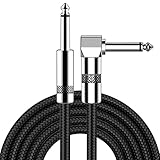
- [Low Noise High Fidelity] New bee guitar cord with 22AWG oxygen-free-copper center conductors provides enhanced signal transfer, the PVC insulation material and the high-density stranded copper double shield provide noise rejection for quiet operation
- [Ideal for Choice] New bee 1/4 straight and right angle metal connectors, ensures a secure connection in any instrument, pedal, or amp jack. L shape great fro on stage guitar stands
- [Wide Compatible] Suitable for mixer, power amplifier, microphone, effector, decoder, equalizer, electoronic organ, electric guitar, electric bass and other electronic equipments. Widely used in the installation of smart home, karaoke speaker, professional stage audio, etc
- [Easy Disassemble] Making incidental repairs quick and easy! Very nice, flexible cable
- [Dont Hesite to Order] 24 hours customer services and professional technology team are standing by
When spatial transcriptomics is the better first choice
Use it when location is part of the hypothesis
Choose spatial data when you need to distinguish a state inside an organ primordium from the same state at a boundary, determine whether a gene gradient follows an anatomical axis, or test whether two populations occupy neighboring domains. The coordinate is the observation that scRNA-seq alone cannot supply.
Use it to validate cell-state assignments in tissue
A cluster discovered by scRNA-seq can be mapped onto a section to test whether its marker genes appear in the expected structure. This can reveal that a putative population is broadly distributed, restricted to a niche, or absent from the region assumed by a dissociation-based interpretation.
Match the assay to the spatial scale
- Broad patterned domains: A spot-based or array approach may provide sufficient coverage when the structures are much larger than a capture feature.
- Small boundaries or adjacent populations: Choose a method whose effective resolution and transcript detection can separate the expected distances; nominal feature size alone is not enough.
- Subcellular localization or very rare cells: Imaging approaches may be appropriate if the required panel, instrumentation, acquisition time, and segmentation quality are available.
Spatial measurements can be sparse. A blank or weak spot may reflect low RNA capture, tissue loss, insufficient sequencing, or true absence. Interpret signal together with morphology, quality metrics, and known markers.
Can you combine scRNA-seq and spatial data for an embryo?
Yes. An integrated design is often the most informative option when you need both unbiased cell-state discovery and anatomical localization. The usual strategy is to use scRNA-seq as a reference atlas and transfer or deconvolve those annotations in spatial data. The reverse is also possible: spatial domains can define subsets for more focused single-cell analysis.
A practical integration sequence
- Generate a reference cell atlas. Profile appropriately staged embryos with scRNA-seq or single-nucleus RNA-seq. Keep biological replicates and batch information explicit.
- Define robust cell states. Use multiple markers and gene programs, not one marker, and check whether states are reproducible across embryos.
- Process the spatial section independently. Perform image quality control, coordinate registration, normalization, and spot or cell segmentation before forcing the single-cell labels onto it.
- Map or deconvolve. Estimate which reference states best explain each spatial feature. For mixed spots, report proportions or uncertainty rather than calling the entire spot one cell type.
- Check the mapping against anatomy. A biologically plausible map should agree with morphology, known landmarks, and independent expression evidence.
- Test spatial patterns. Evaluate whether a state is enriched in a domain, follows a gradient, or forms a boundary, while accounting for section and embryo as sources of variation.
Integration is not a way to recover information that neither assay measured. A mixed spot cannot be turned into a certain single-cell identity merely by applying a reference, and a reference atlas from a different species or developmental stage may map poorly. Reviews of integration methods, including “Integration tools for scRNA-seq data and spatial transcriptomics sequencing data,” emphasize that tools differ in scope and limitations.
Designing an embryo experiment step by step
1. Write the decision question in spatial terms
State whether the primary endpoint is a list of cell states, a developmental transition, a location, a boundary, or a relationship between state and location. If the answer contains both “which cells” and “where,” plan an integrated design from the start rather than adding spatial data after the single-cell experiment is complete.
2. Fix the biological comparison
Specify species, developmental stage, genotype, treatment, sex where relevant, and the number of independent embryos. Do not use technical sections or technical libraries as substitutes for biological replication.
The Tool Desk
Outbyte PC Repair FREERepair Windows errors before they cause bigger problemsFix Now →Outbyte Driver Updater FREEScan for outdated or missing drivers - takes under a minuteDriver Scan →3. Decide what must be resolved
Estimate the size of the structure, boundary, or cell population that matters. Then compare that scale with the assay’s effective resolution, not just the vendor’s nominal feature dimensions. Diffusion, segmentation error, tissue distortion, and mixed-cell capture can enlarge the effective measurement footprint.
Rank #4

- Digital Family Calendar: BSIMB digital calendar wall touch screen puts an end to your chaotic life. Designed for busy families, the digital calendar keeps you organised and in step with your family. Assign different colours and tasks to each family member to make family scheduling easier and more intuitive.
- Easy Setup and Auto-Sync: Plug in, connect to WLAN and auto-syn your electric calendar in minutes. Digital wall calendar compatible with Google calendar, iCloud calendar, Outlook calendar, Cozi calendar and Yahoo calendar. Add events directly on the device or through the ecalendar app. Electronic calendar stays current whether you're at work or traveling—no manual updates needed.
- All-In-One Home Assistant: Smart calendar builds habits and encourage independence with personalised chores. Display daily meal plan to reduce the daily ‘What's for dinner?’ stress. To-do lists keep track of everything you want. Turning chores into valuable achievements with stars and rewards, making life easier and more enjoyable.
- Manage Anytime Anywhere: BSIMB electronic calendar for family stay connected with the powerful ecalendar app. Access and manage your digital wall calendar anytime, anywhere with free features like stars & rewards, photo screensaver, chore charts, meals plans, to-do lists etc. Electronic planner paid features like Magic Import tailor advanced tools for users with special needs.
- Stay in Touch with Loved Ones: Share photos or videos from your phone to electronic wall calendar with app, instantly update your life moment, and keep in touch with your family, especially those who are far away; you can also surprise your smart calendar digital wall by sending him a wish card; it's also great for keeping tech-illiterate elders in touch with the youngsters.
4. Protect orientation and metadata
For spatial work, document cutting plane, anterior-posterior or dorsal-ventral orientation, section order, and landmarks. For scRNA-seq, record any microdissection or regional pooling performed before dissociation. These details determine whether results can be aligned across embryos.
5. Pilot tissue handling
Test fixation, freezing, section thickness, permeabilization, dissociation time, and RNA quality on representative material. A pilot should answer whether morphology survives and whether the expected cell or transcript classes are detectable before committing scarce embryos.
6. Predefine quality and exclusion rules
Set thresholds for low-complexity cells, mitochondrial or stress signatures, doublets, damaged sections, background, and failed segmentation before inspecting biological group labels. Changing rules separately for each condition can create artificial differences.
Quick wins for a faster PC:
Fix the driver behind crashes, sound loss and screen glitchesFind Drivers →Clear out junk files and repair common Windows errorsFree Scan →7. Plan validation
Reserve material or assays for confirming key markers and boundaries. Validation can include targeted in situ detection, imaging, histology, or an independent sequencing preparation, selected to test the specific claim rather than to decorate the final figure.
Independent reader supportYour contribution helps us test, update, and keep practical guides available for everyone.How to interpret results without overclaiming
Separate measurement from inference
A gene count is a measurement; a cluster label is an interpretation; a proposed lineage is a model. State which level each result occupies. In spatial data, a bright signal establishes detected RNA at a coordinate, not automatically the identity or activity of every cell contributing to that coordinate.
Account for sampling
Sections represent selected planes, and scRNA-seq libraries represent captured cells that survived preparation. A rare state may be missed, while a large population may dominate counts. Report the number of embryos, sections, cells, spots, and usable regions so readers can judge coverage.
Use independent evidence for developmental mechanisms
Co-expression and spatial adjacency can suggest a signaling relationship, but they do not prove direct regulation or cell-cell communication. Combine transcript patterns with perturbation, lineage tracing, protein localization, or functional assays when the conclusion concerns mechanism.
Free tools Windows power users keep installed
One-click scans. No signup required.
Keep species and stage boundaries explicit
Embryonic gene programs and anatomy change quickly. A result from E14.0 C57BL/6N mouse embryos should be labeled as such and not presented as a general human-development result. Cross-species or cross-stage comparisons require explicit orthology, staging, and anatomical justification.
Common failure modes and how to fix them
“The spatial map has single-cell resolution, so every spot is one cell.”
Why it fails: Capture features may contain several cells or cell fragments, and diffusion can spread molecules beyond their source. Fix: Use morphology and segmentation, report mixed contributions, and interpret effective rather than nominal resolution.
“The scRNA-seq cluster identifies its original embryonic location.”
Why it fails: Dissociation removes coordinates. Fix: Preserve regional labels before dissociation or map the cluster to spatial data and validate the assignment.
“A larger UMI count means a universally better platform.”
Why it fails: Detection depends on tissue, chemistry, sequencing depth, background, and the spatial area measured. Fix: Compare assays on matched specimens and report the conditions behind the metric.
“One section or one embryo is enough because thousands of spots were measured.”
Why it fails: Spots are not independent biological replicates. Fix: Treat embryo and, where appropriate, section as experimental units and model within-embryo sampling accordingly.
“A failed map means the cell state is absent.”
Why it fails: Poor permeabilization, tissue loss, low depth, background, or an incomplete marker set can erase a real signal. Fix: Inspect quality metrics, morphology, positive controls, and the reference atlas before making an absence claim.
How to choose a spatial platform responsibly
Start by listing the smallest anatomical feature that must be distinguished, the total embryonic area that must fit in one experiment, the number of sections and embryos available, and whether unbiased discovery or a targeted panel is more important. Then compare candidate methods on:
- Effective resolution after diffusion and segmentation, measured in the tissue of interest.
- Transcript detection and gene coverage for the expected embryonic states.
- Field of view and the number of sections that can be processed within the project timeline.
- Compatibility with the required fixation, embedding, and morphology.
- Sequencing depth, imaging time, data storage, and analysis expertise.
- Ability to replicate the assay across embryos and developmental stages.
The 2024 benchmark of 11 sequencing-based methods found method- and tissue-dependent differences, including molecular-diffusion effects on effective resolution. Use such comparisons to formulate a matched pilot, not to transfer a ranking from reference tissues to an embryo automatically. Platform specifications can change, so verify current documentation before ordering an experiment.
A concise decision framework
| If your highest-priority question is… | Start with… | Add or consider… |
|---|---|---|
| Which cell types and transient states exist? | scRNA-seq or single-nucleus RNA-seq | Spatial mapping to verify their anatomical distribution |
| Where is a known state, gradient, or boundary? | Spatial transcriptomics matched to the required scale | scRNA-seq if marker discovery or deconvolution reference is weak |
| How do lineages change and where do they reside? | Integrated scRNA-seq plus spatial analysis | Independent lineage or functional validation |
| Are cells smaller than the assay’s capture feature? | A method with demonstrated effective resolution in the same tissue | Imaging or targeted validation if mixed spots obscure the answer |
| Is the embryo too scarce for separate experiments? | A pilot that prioritizes the decisive measurement | Preserve material for orthogonal validation rather than maximizing one metric |
Bottom line for embryo studies
Use scRNA-seq when cell-state discovery, rare populations, and transcriptional programs are the main objectives. Use spatial transcriptomics when anatomical position, boundaries, gradients, or tissue context are indispensable. Because each method leaves a different blind spot, the strongest developmental studies often combine a single-cell reference with spatial mapping, while stating clearly what the assay’s resolution, sampling, and species-specific evidence can support.
Quick Recap
Product prices and availability are accurate as of the date/time indicated and are subject to change. Any price and availability information displayed on Amazon at the time of purchase will apply.

