Do these 3 things before closing this tab:
1Clear out junk files and repair common Windows errors2Fix the driver behind crashes, sound loss and screen glitches3Repair Windows errors before they cause bigger problemsPSMA PET is the molecular imaging part of the examination. It shows where an injected, radiolabeled ligand binds to prostate-specific membrane antigen (PSMA). PET/CT adds X-ray-based anatomy, while PET/MRI adds magnetic-resonance anatomy. These names therefore describe combinations of one molecular imaging method with different anatomic scans—not three fundamentally different kinds of radiotracer.
The right choice depends on the clinical question, the required anatomic detail, the scan protocol, local expertise and availability, and the patient’s circumstances. Neither PET/CT nor PET/MRI is automatically best for every situation.

As an Amazon Associate I earn from qualifying purchases.
What PSMA PET actually measures
PSMA stands for prostate-specific membrane antigen, a protein found at much higher levels on most prostate cancer cells than on normal cells. The Society of Nuclear Medicine and Molecular Imaging (SNMMI) says prostate cancer cells can have up to 1,000 times more PSMA than normal cells. An injected radiotracer designed to bind PSMA makes those sites visible on a PET scan.
PET creates a three-dimensional map of tracer distribution. Areas of increased uptake may represent prostate cancer, but uptake is not the same as a confirmed diagnosis. Normal organs can show physiologic uptake, and some non-malignant conditions and other cancers can also bind PSMA-targeting agents. A qualified reader must interpret the pattern, intensity and location together with the patient’s history, laboratory results and other imaging.
#1 Best Overall

- [Ultra-Lightweight Ear Buds Designed for Small Ears] Each earbud weighs only 3.7g and features a compact, ergonomic in-ear design made especially for small ears. Secure, low-profile, and comfortable for workouts, all-day wear or overnight sleeping.The ultra-slim profile sits flush in your ear with no hard edges poking or rubbing, so you can even sleep on your side without discomfort.
- [Immersive Stereo Sound with TOZO OrigX Technology] TOZO OrigX tuning delivers clear vocals, balanced mids, and natural stereo sound for music, podcasts, and videos.
- [Long Battery Life for Daily Use] Get up to 7 hours of playtime on a single charge, with up to 32 hours total using the charging case—ideal for workdays, commuting, and extended listening sessions.
- [Bluetooth 5.3 & Stable Connection] Bluetooth 5.3 provides fast pairing, stable wireless performance, and reduced dropouts as you move around home or office.
- [Deep Bass with Clear Vocals] High-performance drivers produce punchy bass while keeping vocals clean and detailed for everyday listening.
Not every prostate tumor expresses PSMA at the same level. A scan can therefore show intense, faint or absent uptake for reasons that require clinical interpretation. A negative finding may reflect a lesion that is too small, has low tracer expression or is affected by technical factors; it is not proof that no cancer exists.
The radiotracer is administered intravenously. Several approved PSMA tracers have broadly similar imaging performance, but approvals and availability vary by country and can change over time. The terms PET, PET/CT and PET/MRI describe the imaging system and anatomy acquired alongside the tracer distribution; they do not, by themselves, identify a unique tracer.
Why the three names are easy to confuse
“PSMA PET” is often used as shorthand for the molecular portion of a study. In practice, a department may schedule that PET acquisition on a PET/CT or PET/MRI scanner. The tracer and PET detector provide the functional information, while the companion CT or MRI supplies anatomic context. Thus, PSMA PET/CT and PSMA PET/MRI are not different PSMA drugs; they are different ways of pairing the same type of molecular examination with anatomy.
What’s actually slowing this PC down?
Pick the symptom - the matching free tool is one click away.
The distinction matters when reading a study or comparing evidence. A result obtained with a low-dose CT for attenuation correction is not equivalent to a result obtained with a full diagnostic CT. Likewise, evidence that adds PSMA PET to a separately acquired multiparametric MRI is not automatically evidence about an integrated whole-body PET/MRI scanner. The indication, acquisition protocol and reference standard must be considered together.
PSMA PET, PSMA PET/CT and PSMA PET/MRI compared
| Term | What it contributes | Important qualification |
|---|---|---|
| PSMA PET | Functional and molecular information: where the PSMA-targeting tracer accumulates. | “PET” alone does not specify which anatomic scan accompanies it. Uptake can be physiologic, benign or related to another cancer. |
| PSMA PET/CT | PET tracer distribution plus structural X-ray images from CT. CT also supports PET attenuation correction and helps localize uptake. | The CT may be a low-dose acquisition for attenuation correction and correlation, not a full diagnostic CT. The protocol must be confirmed in the report or with the imaging department. |
| PSMA PET/MRI | PET tracer distribution plus MRI-derived anatomy. PET and MRI may be acquired sequentially or simultaneously while the patient remains positioned in the integrated system. | Applications have been reported, including initial staging and biochemical recurrence, but the 2023 joint EANM/SNMMI procedure guideline specifically excludes PET/MRI protocols from its scope. |
How CT and MRI change the information you get
What CT adds
CT provides cross-sectional X-ray anatomy. It can show the size and shape of structures, identify bones and many soft-tissue abnormalities, and place a focus of PET uptake in an anatomic location. In a PET/CT examination, CT data are also used to correct for attenuation of the PET signal.
“PET/CT” does not always mean that a comprehensive, contrast-enhanced diagnostic CT was performed. Many examinations use a low-dose CT primarily for attenuation correction and localization. If a detailed diagnostic CT is clinically necessary, it may need to be ordered or performed as a separate protocol. Ask specifically whether intravenous contrast, diagnostic-quality images or only a low-dose localization CT is planned.
What MRI adds
MRI supplies anatomy using magnetic fields and radiofrequency signals rather than X-rays. Multiparametric prostate MRI can characterize the prostate and surrounding tissues with sequences that provide different structural and functional contrasts. Whole-body or regional MRI can also contribute soft-tissue and marrow information, depending on the protocol.
The Tool Desk
Outbyte PC Repair FREERepair Windows errors before they cause bigger problemsFix Now →Outbyte Driver Updater FREEFix the driver behind crashes, sound loss and screen glitchesFind Drivers →PET/MRI can be an integrated or multimodality system. PET and MRI may be acquired one after the other or simultaneously, with the patient remaining in the same position. That arrangement can help align the two datasets, but it does not by itself guarantee better diagnostic performance. The quality of the MRI sequences, motion control, field of view, contrast decisions and reader expertise all affect the result.
The fact that MRI offers particular soft-tissue strengths does not establish that PET/MRI is universally more accurate, safer or preferable. Its value depends on the body region, protocol quality, patient suitability and the expertise available to interpret both datasets.
Rank #2

- 【IP68 Waterproof Multifunctinal Smart Watch】Torjalph fitness activity tracker smart watch is IP68 waterproof technology ,you can wear it when swimming,handwashing,it's rain-proof which means that you perform multiple exercise modes outside without worrying about sweat or rain corroding your fitness smart watch,and this watch equipped with a 230 mAh large capacity battery magnetic charging ,and this sports activity tracker watch can be used 5-7 days If fully charged.
- 【Larger 1.83'' Full Screen Watch & Multi-dial Selection】1.83 inch full HD color touch screen smart watch for men / women provides excellent 240*284 resolution image quality ,This Bluetooth smart watch offers a better touch experience and remarkable immersion and ,more than 200 watch faces for you to choose from App"Da Fit ", you can change the dial style according to your mood to wear keeping the world on your wrist
- 【Accurate Activity Tracker Smart Watch 】sports watch activity tracker uses sweat and dust resistant breathable silicone strap, our Fitness Tracker Smart Watch for Android iOS Phones supports 12 sports modes,walking,running,bicycle,swimming,badminton,basketball,football,rope skipping. This bluetooth calls smart watch for men women is lightweight and stylish, bringing more convenient and relaxing sports experience while also providing more protection.
- 【Your 24 Hour Personal Daily Health Monitor】Fitness watch for women men full touch screen IP67 waterproof bluetooth smartwatch for Android iOS Phones with heart rate blood pressure monitor multifunction Andriod smart watch with steps counter sleep monitor sports fitness watch, monitoring blood oxygen saturation allows you to understand your physical condition in real time to track fitness progress ,to know your health status in time.
- 【More Functions Bluetooth Call Smart Watch】This fitness activity smart watch full screen for iPhone and Android phones includes a bunch of new features including:The bluetooth smart watch can hang up the phone, receive and display WhatsAPP /Text /Facebook /Twitter /Messenger reminder.Support sedentary reminder, alarm clock, stopwatch, timer,weather information,music control, remote camera control compatible with Android 5.0 and above or iOS 9.0 and above smartphones.
When PSMA PET/CT is used
The joint European Association of Nuclear Medicine (EANM) and SNMMI procedure guideline, version 2.0 (2023), describes several routine clinical questions for PSMA-ligand PET/CT. These are indication-based uses, not a recommendation to scan everyone with prostate cancer or everyone with an elevated PSA.
Initial staging
For people with newly diagnosed prostate cancer, PSMA PET/CT can help assess whether disease extends beyond the prostate, including lymph nodes or distant sites. In the phase III multicenter randomized ProPSMA trial, gallium-68 PSMA-11 PET/CT had 27% greater accuracy than conventional CT plus bone scanning for initial staging of people with high-risk prostate cancer. That percentage applies to the trial population and its specific comparison; it is not a universal estimate for PET/CT versus PET/MRI or for every risk group.
The result also does not mean that every suspicious focus is malignant or that a negative scan excludes microscopic disease. Treatment planning still incorporates biopsy grade, PSA, examination findings and other clinical information.
Persistent or recurrent disease
When prostate-specific antigen (PSA) remains detectable after treatment or rises later, PSMA PET/CT can help localize disease that conventional imaging has not shown. The scan may identify a site in the prostate bed, lymph nodes or distant organs, allowing the clinical team to consider targeted evaluation or treatment. The significance of a finding depends on the timing of treatment, PSA trend, prior radiation or surgery and whether the location is technically amenable to intervention.
Selected conventionally non-metastatic castration-resistant disease
The guideline includes localization in selected patients whose disease is considered non-metastatic on conventional imaging but is progressing despite androgen-deprivation treatment. “Selected” matters: eligibility and interpretation depend on the clinical record and the imaging question, not simply on a PSA value. A scan should be ordered through the treating team, which can determine whether the result would change management.
Staging before PSMA-directed radioligand therapy
PSMA PET/CT can be used to establish disease distribution and PSMA expression before a PSMA-directed radioligand treatment is considered. Treatment programs apply their own imaging and eligibility criteria. A scan intended for therapy selection may use a different reporting focus from a scan intended to investigate an isolated PSA rise.
Free tools Windows power users keep installed
One-click scans. No signup required.
Other and emerging applications
The guideline also discusses possible roles in biopsy guidance and imaging of metastatic disease, while noting uncertainty for some developing uses. PSMA PET/CT is not a population screening test for people without a clinical indication. The fact that a scanner can detect tracer uptake does not mean it should be used as a routine check in people with no prostate-cancer concern.
What is known about PET/MRI
Published work has reported PET/MRI use for areas such as initial staging and biochemical recurrence. However, the joint EANM/SNMMI guideline addresses PET/CT procedure protocols and expressly states that PET/MRI protocols are outside its scope. That means the guideline cannot be used to claim a settled PET/MRI protocol or a universal superiority claim.
A key distinction is often missed: evidence that combines PSMA PET with multiparametric prostate MRI is not automatically evidence that an integrated whole-body PSMA PET/MRI scanner is better than PSMA PET/CT. The clinical setting, acquisition protocol and reference standard must match before results can be compared.
Rank #3
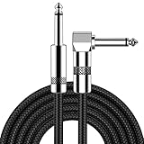
- [Low Noise High Fidelity] New bee guitar cord with 22AWG oxygen-free-copper center conductors provides enhanced signal transfer, the PVC insulation material and the high-density stranded copper double shield provide noise rejection for quiet operation
- [Ideal for Choice] New bee 1/4 straight and right angle metal connectors, ensures a secure connection in any instrument, pedal, or amp jack. L shape great fro on stage guitar stands
- [Wide Compatible] Suitable for mixer, power amplifier, microphone, effector, decoder, equalizer, electoronic organ, electric guitar, electric bass and other electronic equipments. Widely used in the installation of smart home, karaoke speaker, professional stage audio, etc
- [Easy Disassemble] Making incidental repairs quick and easy! Very nice, flexible cable
- [Dont Hesite to Order] 24 hours customer services and professional technology team are standing by
PET/MRI may be considered where anatomic MRI detail is particularly important and the center has appropriate equipment and expertise. It may be less suitable for a person with a non-compatible implanted device, substantial claustrophobia or another MRI-specific limitation. These practical issues are part of modality selection, not evidence that one system wins in every patient.
Evidence that helps put the numbers in context
Pelvic lymph-node disease
Across two phase II/III studies summarized by the EANM/SNMMI guideline, pelvic-node specificity was at least 95%, while sensitivity was in the 40% range. High specificity means a convincing positive finding can be useful for confirming disease in the right context. Sensitivity in that range means microscopic nodal metastases are frequently too small to detect. A negative PSMA PET scan therefore cannot exclude every pelvic lymph-node micrometastasis.
This is why a surgeon or oncologist may still discuss pelvic treatment or lymph-node assessment despite a negative scan. The scan answers a probability question; it does not replace pathology when tissue confirmation is required and feasible.
Biopsy guidance: the PRIMARY study
In the prospective PRIMARY study, adding gallium-68 PSMA-11 PET to multiparametric MRI increased the negative predictive value for clinically significant prostate cancer from 72% to 91% and sensitivity from 83% to 97%. The guideline uses these findings to support combining PSMA PET with multiparametric MRI for biopsy guidance. They do not prove that PET/MRI is always superior to PET/CT, because the study evaluated an added combination in a particular biopsy setting.
Negative predictive value depends on the population being tested and the definition of clinically significant disease. The PRIMARY percentages should therefore not be transplanted to every PSA level, biopsy strategy or scanner configuration.
Pylarify PET/CT after treatment
In a 208-patient study cited by the U.S. Food and Drug Administration (FDA), participants had rising PSA after definitive treatment and baseline conventional imaging that did not show definite spread. Pylarify PET/CT detected at least one positive lesion in 60% of the study cohort. Among patients with correlative pathology, imaging or serial PSA information, the FDA reported confirmation of positive readings in an estimated 85% to 87%, depending on the reader. These figures belong to that cohort and verification process; they are not a general detection rate for all PSMA PET examinations.
What happens during a typical PSMA PET examination
Exact preparation and timing vary by tracer, facility and whether CT or MRI is included. SNMMI’s patient guide describes a typical workflow as follows:
- Check the protocol and safety requirements. Tell the imaging team about pregnancy or possible pregnancy, breastfeeding, kidney problems, allergies, implanted devices and prior imaging reactions. MRI adds device-compatibility and metal-safety screening. Ask whether contrast is planned and whether any additional preparation is required.
- Receive the tracer through an IV. The radiotracer is administered in a controlled clinical setting. It is not a consumer product and cannot be self-administered. Staff verify the order, the patient and the tracer before injection.
- Wait for distribution. The patient guide estimates a wait of about one hour after injection. Local protocols can be shorter or longer. The waiting period allows the tracer to circulate and clear from tissues before imaging.
- Complete the scan. The guide estimates about 30 minutes of scanning. PET/CT and PET/MRI times differ according to the anatomic coverage and whether additional diagnostic sequences are obtained. Remaining still is important, particularly during MRI sequences.
- Have the images interpreted. A qualified imaging specialist reviews PET findings with CT or MRI anatomy and the available clinical information, then issues a report for the treating team. The ordering clinician explains how the result affects the next decision.
Patients should not assume that the injection time or scanner time is the total appointment length. Registration, safety screening, IV placement, uptake time and any additional MRI or diagnostic CT sequences can extend the visit.
Limitations, false results and safety context
A negative scan is not a guarantee
Very small tumors may fall below PET’s spatial resolution. SNMMI’s patient information gives tumors smaller than about 0.5 cm as an example that may not appear. The pelvic-node sensitivity data provide a second, disease-specific reason a negative scan cannot rule out microscopic spread.
Rank #4

- Digital Family Calendar: BSIMB digital calendar wall touch screen puts an end to your chaotic life. Designed for busy families, the digital calendar keeps you organised and in step with your family. Assign different colours and tasks to each family member to make family scheduling easier and more intuitive.
- Easy Setup and Auto-Sync: Plug in, connect to WLAN and auto-syn your electric calendar in minutes. Digital wall calendar compatible with Google calendar, iCloud calendar, Outlook calendar, Cozi calendar and Yahoo calendar. Add events directly on the device or through the ecalendar app. Electronic calendar stays current whether you're at work or traveling—no manual updates needed.
- All-In-One Home Assistant: Smart calendar builds habits and encourage independence with personalised chores. Display daily meal plan to reduce the daily ‘What's for dinner?’ stress. To-do lists keep track of everything you want. Turning chores into valuable achievements with stars and rewards, making life easier and more enjoyable.
- Manage Anytime Anywhere: BSIMB electronic calendar for family stay connected with the powerful ecalendar app. Access and manage your digital wall calendar anytime, anywhere with free features like stars & rewards, photo screensaver, chore charts, meals plans, to-do lists etc. Electronic planner paid features like Magic Import tailor advanced tools for users with special needs.
- Stay in Touch with Loved Ones: Share photos or videos from your phone to electronic wall calendar with app, instantly update your life moment, and keep in touch with your family, especially those who are far away; you can also surprise your smart calendar digital wall by sending him a wish card; it's also great for keeping tech-illiterate elders in touch with the youngsters.
A negative result should be read alongside PSA behavior, pathology, examination findings and prior imaging. If clinical suspicion remains high, the treating team may recommend follow-up imaging, biopsy or another test rather than treating the negative scan as definitive.
Positive uptake is not synonymous with prostate cancer
Normal organs have expected physiologic uptake, and benign processes or other cancers can bind PSMA-targeting agents. The FDA warns that Pylarify binding in other cancers and some non-malignant conditions can lead to interpretation errors. Location, pattern, intensity and comparison with other studies all matter.
For an unusual focus, the report may recommend correlation with diagnostic CT, MRI, laboratory data, follow-up imaging or tissue sampling. A radiology phrase such as “equivocal” reflects uncertainty that must be resolved in clinical context; it is not an automatic cancer diagnosis.
Protocol differences affect comparisons
A low-dose CT for attenuation correction is not interchangeable with a diagnostic CT. MRI protocols also vary in field of view, sequences, contrast use and motion control. A result from one protocol should not be generalized to every PET/CT or PET/MRI examination.
Quick wins for a faster PC:
Scan for outdated or missing drivers - takes under a minuteDriver Scan →Clear out junk files and repair common Windows errorsFree Scan →Fix the driver behind crashes, sound loss and screen glitchesFind Drivers →Even the same tracer can produce different practical results when uptake time, injected activity, scanner technology, reconstruction settings or reader experience differ. These details are reasons to compare like with like when discussing published accuracy figures.
Radiation and other practical considerations
PET/CT includes radiation from the radiotracer and CT. PET/MRI avoids CT’s X-ray exposure but still involves the radiotracer and may be unsuitable for some implanted devices, severe claustrophobia or other MRI-specific reasons. The imaging team weighs these factors against the information needed.
As SNMMI states in its patient guidance, “No imaging test is perfect.” The report must be interpreted alongside PSA trends, examination findings, pathology and prior imaging rather than treated as a stand-alone diagnosis.
Independent reader supportYour contribution helps us test, update, and keep practical guides available for everyone.How clinicians decide between PET/CT and PET/MRI
There is no single modality ranking that applies to every patient. A useful decision starts with the question the scan must answer.
Recommended Free Tools
| Decision factor | Why it matters | What to clarify |
|---|---|---|
| Clinical question | Initial staging, recurrence localization, biopsy planning and treatment eligibility require different anatomic detail. | Ask what decision the scan is intended to change. |
| Anatomic information needed | CT and MRI depict anatomy differently; MRI may be particularly useful when detailed soft-tissue or prostate characterization is required. | Ask whether multiparametric MRI is needed in addition to PET and whether it is integrated or performed separately. |
| CT protocol | A low-dose CT for attenuation correction and localization does not necessarily answer every diagnostic CT question. | Confirm whether contrast or a full diagnostic CT protocol is included. |
| Patient suitability | MRI requires screening for implants, metal and other contraindications; CT and radiotracer decisions have their own safety considerations. | Provide a complete implant, allergy, kidney, pregnancy and breastfeeding history. |
| Local expertise and access | Image quality, reporting experience, scheduling and tracer availability differ among centers. | Ask who interprets the study and which protocol the center routinely uses for your indication. |
| Evidence limits | Evidence for PSMA PET/CT indications is more explicitly covered by the joint guideline than PET/MRI protocols. | Request the rationale for the chosen modality rather than assuming one is universally better. |
In some cases, the choice is not either/or. A clinician may request PSMA PET/CT for whole-body staging and a separate multiparametric MRI for detailed prostate assessment. In another case, an integrated PET/MRI examination may be available and clinically reasonable. The ordering team should explain whether the studies answer the same question or complementary questions.
Questions to ask before receiving a result
- Which PSMA-targeting tracer will be used, and is it approved or available for this indication in this region?
- Is the CT low-dose for attenuation correction, or is a diagnostic CT protocol included?
- If MRI is used, is it multiparametric prostate MRI, whole-body MRI, or another protocol?
- What clinical question is the scan intended to answer?
- Could a negative result miss microscopic lymph-node or very small-volume disease?
- Could a positive focus have a benign or non-prostate-cancer explanation?
- How will the result be combined with PSA, pathology, examination findings and previous imaging?
- Who will interpret the PET component and the CT or MRI component?
- Will the scan be used to plan biopsy, surgery, radiation or radioligand therapy?
- Are there MRI implant, claustrophobia, kidney, allergy, pregnancy or breastfeeding issues the center needs to know about?
Bottom line
PSMA PET is the tracer-based molecular map. PSMA PET/CT adds CT anatomy, and PSMA PET/MRI adds MRI anatomy. PSMA PET/CT has established guideline-supported uses for several prostate cancer staging, recurrence and treatment-planning questions, with important limits for microscopic disease and nonspecific uptake. PET/MRI is a reported option in some settings, but the joint EANM/SNMMI guideline does not establish its protocols or make it a universal replacement for PET/CT. The treating team and qualified imaging specialists should select and interpret the examination for the specific clinical question.
Frequently Asked Questions
Is PSMA PET the same as PSMA PET/CT?
PSMA PET describes the molecular scan that maps an injected PSMA-targeting tracer. PSMA PET/CT adds CT anatomy to that PET information. The terms do not describe different tracers.
Is PSMA PET/MRI better than PSMA PET/CT?
Not universally. MRI and CT provide different anatomic information, and the joint 2023 EANM/SNMMI guideline covers PET/CT procedures while excluding PET/MRI protocols. The appropriate choice depends on the clinical question, protocol, patient suitability and local expertise.
Windows Errors? Fix Them Before They Spread
Repair common Windows errors and clear accumulated junk for a smoother, more stable PC - no reinstall needed.Free scan · no reinstallOutdated Drivers Are Slowing You Down
One free scan finds every outdated or missing driver and matches the right update for your exact hardware.Free scan · exact hardware matchCan a negative PSMA PET scan rule out prostate cancer?
No. Very small tumors, including lesions smaller than about 0.5 cm, may not appear, and pelvic lymph-node sensitivity has been reported in the 40% range. A negative scan must be interpreted with PSA, pathology and other findings.
Does PSMA uptake prove that a spot is prostate cancer?
No. Normal organs have physiologic uptake, and some benign conditions and other cancers can bind PSMA-targeting agents. The pattern and location require interpretation by a qualified imaging specialist.
How long does a typical PSMA PET appointment take?
SNMMI’s patient guide estimates about one hour between injection and imaging and about 30 minutes of scanning. Actual appointment times vary with the tracer, anatomic coverage, CT or MRI protocol and any additional sequences.
What should I tell the imaging center before PSMA PET/CT or PET/MRI?
Report pregnancy or possible pregnancy, breastfeeding, kidney problems, allergies, implanted devices, prior imaging reactions and, for MRI, any metal or device-compatibility concerns. Ask whether the CT is low-dose or diagnostic and which MRI protocol is planned.
Free tools Windows power users keep installed
One-click scans. No signup required.
The Bottom Line
PSMA PET supplies molecular information; CT or MRI supplies anatomy. PET/CT has the clearest established guideline role, while PET/MRI is used in selected settings without a universal superiority claim. Scan selection and interpretation belong with the treating team.
Quick Recap
Product prices and availability are accurate as of the date/time indicated and are subject to change. Any price and availability information displayed on Amazon at the time of purchase will apply.

