Free tools Windows power users keep installed
One-click scans. No signup required.
A nanoparticle platform called CMBFO made tumors visible on MRI and generated reactive oxygen species (ROS) by two routes in mice carrying subcutaneous tumors derived from U87 glioma cells. The work is preclinical. It does not show a treatment for glioma in people, and the mice did not have brain tumors.
What CMBFO is
CMBFO stands for chitosan-functionalized, manganese-doped bismuth ferrite nanoparticles. The authors describe it as an experimental nanoplatform. It is not a licensed drug, a device, or a product available to patients or clinicians. Its three intended functions are to act as a pH-responsive contrast agent for T2-weighted MRI, to produce ROS through chemodynamic therapy (CDT), and to produce ROS through ultrasound-activated piezodynamic therapy (PZDT). The study is reported by Gong et al. in the Journal of Nanobiotechnology (2026).
As an Amazon Associate I earn from qualifying purchases.
How the three functions work
MRI signal tied to acidity
Chitosan is a natural polymer whose amino groups pick up protons in acidic conditions. The authors describe tumor-microenvironment-like acidity as causing those groups to become protonated, which loosens the chitosan coating and allows manganese ions (Mn2+) to be released. Released manganese shortens T2 relaxation and so brightens the T2-weighted image, which is the basis of the MRI monitoring. In the primary abstract, the pH-responsive MRI signal peaked at pH 5.1 and six hours after injection.
Chemodynamic ROS generation
Once manganese is released, the authors propose a Fenton-like reaction that converts hydrogen peroxide present in the tumor environment into hydroxyl radicals. This is CDT. Because it depends on the acidic environment that releases the manganese, it is intended to continue as long as that chemistry is active, rather than being a single burst.
#1 Best Overall

- Life Size: Life size human skull and brain model allow you clearly see all the main anatomical structures of the human skull and brain, perfect for patient education or anatomical study.
- 11 Part: The skull model consists of 3 part. The brain model consists of 8 components: sagittal section of brain, cerebral hemisphere, cerebellum and brainstem.
- Numeric Marker: The brain model features a labeled diagram for 32 numeric marker. The skull model includes a labeled diagram for 55 numeric marker.
- Material: Made of robust PVC material. Light-weight, high strength, anti-corrosion, long term service.
Ultrasound-triggered piezodynamic ROS generation
Bismuth ferrite is piezoelectric, meaning it generates charge under mechanical stress. When ultrasound is applied, the material is proposed to catalyze ROS production, which the authors call piezodynamic therapy. The design goal is to pair this fast, externally controlled effect with the slower, acid-dependent chemistry of CDT.
| Feature | Chemodynamic therapy (CDT) | Piezodynamic therapy (PZDT) |
|---|---|---|
| Trigger | Acidity releases Mn2+ from the chitosan coating | Ultrasound applied to the tumor |
| Proposed ROS mechanism | Fenton-like reaction with tumor hydrogen peroxide | Piezocatalysis in the bismuth ferrite particles |
| Proposed timing | Sustained while acidic conditions persist | Rapid, controlled by when ultrasound is applied |
| Endpoint reported in the primary abstract | Not reported separately | Over 95% tumor-cell lethality under ultrasound (cell-lethality measure) |
The two modes are presented as complementary within one particle. The paper does not report a head-to-head comparison of CDT alone against PZDT alone, so the table should not be read as a ranking of the two.
Rank #2
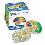
- HELP your child understand the complexities of the human brain with this labeled cross-section model
- EXPLORE the human brain through hands-on science investigation
- Labeled cross-sections of the brain are realistically detailed
- Features the main parts of the brain including - frontal lobe, medulla oblongata, thalamus, cerebellum, hypothalamus, and more
- GIVE THE GIFT OF LEARNING: Whether you’re shopping for holidays, birthdays, or just because, toys from Learning Resources help you discover new learning fun every time you give a gift! Ideal gift for Halloween, Christmas, Stocking Stuffers, Easter or even for Homeschool.
The numbers, and who reported them
Several figures have circulated in coverage of this study. They come from different sources and measure different things, so they should not be combined into one efficacy claim.
The Tool Desk
Outbyte Driver Updater FREEScan for outdated or missing drivers - takes under a minuteDriver Scan →Outbyte PC Repair FREEClear out junk files and repair common Windows errorsFree Scan →| Figure | What it measures | Source and status |
|---|---|---|
| MRI signal peak at pH 5.1 and six hours after injection | Timing and acidity dependence of the pH-responsive MRI signal | Primary abstract, Gong et al., Journal of Nanobiotechnology, 2026 |
| Over 95% tumor-cell lethality under ultrasound | Cell death in the study’s evaluation under ultrasound; a cell-lethality measure, not a tumor-volume percentage | Primary abstract, Gong et al., 2026 |
| About 95% reduction in tumor volume within 12 days | Tumor volume change | Secondary news coverage of the paper; not verified against the primary paper’s figure-level results |
| 38% transport ratio | Transport across an in-vitro blood-brain barrier (BBB) model | AZoNano report, 2026; secondary reporting, not a living-animal measurement |
| About 60% rise in tumor MRI signal-to-noise ratio | Imaging signal in the tumor model | AZoNano report, 2026; secondary reporting |
| Over 80% survival at day 30 | Survival in one treatment group of five mice | AZoNano report, 2026; secondary reporting; group size is very small |
Until these secondary figures are confirmed against the paper’s own figures, treat the volume and survival numbers as reported claims rather than established results. The 95% cell-lethality figure is the only efficacy number taken directly from the primary abstract.
Rank #3

- 【2X Life-Size Brain Model】This medical-grade brain model magnifies 130 key structures at 2X scale, with 4 magnetic detachable sections highlighting emotion, memory, and learning centers. Perfect for Schools, hospitals, visual aids in physical health teaching.Our brain model anatomy can be used in therapy practices or college anatomy class to teach clients or study what's happening in parts of the brain during stress, anxiety, panic, emotions, memories, etc
- 【Colored Design and Laser-Engraved Labels】The brain model for neuroscience consists of nine vividly colored sections to easily distinguish between the different brain structures, that resist fading, blurring, or smudging.Unlike the hand-written labels used by other living human brain model that fade over time, even without cleaning.
- 【Healty Material Quality】The brain anatomy model is made of green and washable PVC material. Durable, Easy to clean.Due to closed space shippment, there may be some smell, but smell will fade away after putting in open air.
- 【4 Magnetic Detachable Sections】The human brain model is double-life-size and consists of 4 parts that are easy to detach and reassemble. This allows for comprehensive neuroanatomy study of the cerebral hemispheres, brain stem and cerebellum. The 4 detachable parts are held together using magnets making it the safest model for even kids to use and study.
- 【Wide Application】 Our realistic human brain anatomical model 2-piece colour coded can as an ideal study tool for medical anatomy teachers or students, and children's interest development learning tool, which can be used in teaching explanation, doctor-patient communication, model display, learning research or laboratory exhibition.
Why the mouse model matters
Subcutaneous tumors are not brain tumors
The primary paper describes tumors grown under the skin of mice. Glioma is a tumor of the brain and spinal cord, and the brain differs from subcutaneous tissue in blood supply, immune environment, and how drugs reach the tumor. A result in subcutaneous U87-derived tumors shows that the particles behave as designed in that setting. It does not show how they would behave in a tumor inside the skull.
Blood-brain barrier data come from a lab model
Brain tumors must be reached through the blood-brain barrier, and news coverage highlighted the 38% transport figure for that reason. That figure comes from an in-vitro barrier model. No living intracranial glioma model is reported as having tested it, so transport across a laboratory barrier is not evidence that the particles would reach a tumor in a brain.
Rank #4

- EMPOWER, UPLIFT, and FEEL GOOD: This adorable, huggable plush brain makes a thoughtful gift for students, educators, healthcare professionals, and anyone passionate about brain health, psychology, and neuroscience. Use it alongside neuroscience workbooks, psychology courses, and classroom lessons or include it in mental health care packages, surgery care packages, medical school gift baskets and more. Pair it with our other brain bestsellers like Brain Cell, Brain Science Themed Keychain Set, Anxiety and more to build the ultimate neuroscience-inspired collection.
- INTERACTIVE ANATOMY EDUCATION: Meet the brain plush that actually teaches - this 6.5" x 5" x 4" brain features embroidered details of the cerebrum, cerebellum, and brainstem for hands-on neuroscience exploration. Designed with medical advisors for anatomical reference, it includes a fact-filled educational card covering brain anatomy, neuron communication, memory, learning, and the incredible ways the brain controls the body. Perfect for classrooms, STEM education, neurology offices, psychology practices, anatomy lessons, and anyone who learns better by holding the lesson in their hands.
- PREMIUM QUALITY and DESIGN: Since 2002, GIANTmicrobes has been developing high quality plush health and biology products. Every parent and gift-giver can shop with confidence. All-new polyester plush materials are non-toxic and tested to ASTM F963 toy safety standards, holds a CPSC Children’s Product Certificate backed by third-party testing and maintains REACH and Prop 65 safety standards. Every design is rooted in real anatomy because health education should be approachable, accurate, and yes – huggable! Care is Simple: surface clean only with a damp cloth - do not machine wash
- COMFORT COMPANION FOR RECOVERY AND BEYOND: Whether it’s brain surgery recovery, a brain injury awareness moment, or any brain health or mental health journey, this brain gift delivers genuine laughter and warmth when it matters most. More meaningful than a standard plush toy, it’s designed to connect with what someone is experiencing, offering comfort and encouragement. The ultra-soft polyester plush exterior wraps a medium-density polyester fiber fill that holds its shape through frequent hugging and squeezing - making it a cozy companion for hospital stays and recovery at home alike.
- UNFORGETTABLE FOR HEALTHCARE HEROES: Show some love to the medical heroes who keep things running smoothly! Makes thoughtful gifts for psychologists, gifts for scientists, medical gifts, medical student gifts, neuroscience gifts, psychology gifts, neurologist gifts, and neurosurgeon gifts. Ideal for Nurses Week, Mental Health Month, brain awareness events, or any just-because appreciation moment that deserves something genuinely memorable.
What the study does not establish
- That CMBFO treats glioma or any brain tumor in animals with intracranial disease.
- That the 95% cell-lethality measure equals tumor shrinkage, survival benefit, or cure.
- That the secondary tumor-volume and survival figures match the paper’s own data.
- Long-term safety, toxicity, or clearance of manganese and bismuth ferrite particles.
- Efficacy, dosing, or safety in people, or any timeline for clinical availability.
Publication status and approvals
The primary article page identifies the paper as an accepted early version published on 1 October 2026. Springer Nature states that it is citable but may be further edited before it is replaced by the final Version of Record, so wording and figures may change. The page also reports approval for the animal work from the Shenzhen University Institutional Animal Care and Use Committee under protocol IACUC-202400090.
How to read future coverage of this platform
- Check the tumor model first. Confirm whether the tumors were subcutaneous or intracranial and whether they were U87-derived.
- Match each number to its endpoint. Cell lethality, tumor volume, imaging signal, and survival are different measurements.
- Check the sample size and the source of each figure, especially for group sizes of a few animals.
- Separate in-vitro barrier data from in-vivo results.
- Look for the final Version of Record before relying on specific values.
Experimental nanoplatforms like this one often move through cell studies, animal studies, and larger safety testing before any human trial, and this paper sits at the animal stage.
Quick Recap
Product prices and availability are accurate as of the date/time indicated and are subject to change. Any price and availability information displayed on Amazon at the time of purchase will apply.

