Nanofibre scaffolds do not instantly reconnect nerves. They are experimental, implantable sheets or conduits designed to guide nerve growth across or around an injury. The evidence described here comes chiefly from rat studies; it does not establish a treatment proven in people or a device available to patients.
What “reconnecting” a nerve means
Peripheral nerves carry signals involved in movement and sensation. When one is injured, a nerve-guidance scaffold may be placed at the site to support regenerating nerve fibres. Depending on its design, it can provide physical direction, carry a biological cue, or release a compound locally.
As an Amazon Associate I earn from qualifying purchases.
These approaches are not one interchangeable treatment. A crush injury, in which the nerve remains continuous, differs from a nerve gap that a conduit must bridge. Results also depend on the animal model, scaffold material and geometry, added cues, comparator, follow-up time, and whether researchers measured tissue structure, nerve conduction, or function.
The Tool Desk
Outbyte PC Repair FREEClear out junk files and repair common Windows errorsFree Scan →Outbyte Driver Updater FREEScan for outdated or missing drivers - takes under a minuteDriver Scan →What the studies have found
Methylcobalamin-releasing sheet in a rat crush model
In 2017, Osaka University described an electrospun biodegradable sheet containing methylcobalamin (MeCbl), a form of vitamin B12, for local implantation at an injured nerve. The sheet released MeCbl for at least eight weeks in an in-vitro laboratory test. In a rat sciatic-nerve crush model, the report described motor, sensory, nerve-conduction, and myelination outcomes. This is evidence about an implanted material in an animal model—not evidence that oral B12 supplements reconnect nerves. Osaka University’s report
#1 Best Overall

- 3000X Enlarged Neuron Anatomy Model: This highly detailed neuron model is enlarged approximately 3000 times, allowing clear visualization of microscopic neural structures that are difficult to observe in standard anatomy resources. Ideal for demonstrating the organization of nerve cells and neural pathways.
- Comprehensive Neuron Structure Display: Accurately illustrates the major components of a neuron, including the cell body (soma), nucleus, dendrites, axon, myelin sheath, Schwann cells, nodes of Ranvier, and axon terminals. Provides a complete overview of neuronal anatomy.
- Detailed Myelinated Nerve Fiber Representation: Features enlarged cross-sectional and longitudinal views of myelinated nerve fibers, helping students understand nerve impulse conduction, insulation provided by myelin, and the relationship between neurons and supporting cells.
- Color-Coded Design for Enhanced Learning: Different anatomical structures are highlighted with distinct colors for quick identification and improved comprehension. The visually engaging design supports effective classroom instruction, laboratory demonstrations, and self-study.
- Ideal for Biology and Medical Education: Suitable for neuroscience courses, biology classrooms, anatomy laboratories, nursing programs, medical schools, and healthcare training environments. A valuable teaching aid for explaining nervous system structure and function.
Soft mesh wrapped around an injured nerve
A separate 2017 announcement from Japan’s National Institute for Materials Science (NIMS) described a very soft mesh made from biodegradable plastic, with fibres several hundred nanometres in diameter. It reported rat sciatic-nerve regeneration and motor and sensory recovery, with recovery described as occurring within six weeks. The announcement said clinical application was being considered at the time; that historical statement does not establish current approval or availability. Osaka University’s account of the related work describes axon regeneration at six months after surgery, a different reported timeline. These accounts should not be collapsed into one follow-up result. NIMS’s announcement
Aligned fibres and matrix hydrogel in a defined nerve gap
A 2021 rat study tested electrospun poly(L-lactic acid) (PLLA) conduits in a 5 mm sciatic-nerve defect. It compared randomly oriented and longitudinally aligned fibres, with and without a hydrogel made from porcine decellularized nerve matrix (pDNM), and included an autograft comparator.
Rank #2
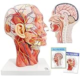
- Non-toxic environmentally friendly PVC material, easy to clean. This head model is 360 degree rotable for easy teaching or self-learning at home.
- This anatomical model of the head shows the local morphology of the inner and outer sides of the mid-sagittal section of the head and neck and its blood vessels and nerves: including the superficial muscles of the exposed face, superficial blood vessels and nerves of the face and scalp And the medial structure of the head and brain and upper airway and the sagittal cross-sectional structure of the cervical spine.
- It is also provided with a detailed product manual with all anatomical structures marked and described with text, which is convenient for teaching.
- This model is the first choice for teaching in classroom or at-home for human head and brain anatomy education, or beauty salons or hosipital & clinc
- Anatomical Features: Ear is detachable to observe external acoustic pore, this model also shows all surperficial face muscles, blood vessel and nerves, parotid gland, submandular gland, medial sagittal section of brain, nasal cavity, oral cavity, larynx and pharynx, tongue, and vertebrae & spinal cord at neck region
Among the conduit variants described, the aligned PLLA conduit with 0.25% hydrogel produced the strongest results, including a sciatic functional index (SFI) comparable to the autograft group eight weeks after surgery. The autograft group nevertheless had the highest SFI across the reported two-to-eight-week period. These are findings in rats, not evidence of equivalent outcomes in people. The 2021 study in Theranostics
Recommended Free Tools
Other experimental scaffold designs
A 2012 study abstract describes a self-assembling nanofibre scaffold inside a blood-vessel conduit used to repair a 10 mm rat sciatic-nerve gap. It reports axon regeneration across and beyond the gap, along with improvements in measures including reinnervation and functional recovery. This was a different animal experiment and scaffold design from the later PLLA study. The PubMed record
Rank #3

- HIGH QUALITY MATERIAL - The model is made of polyvinyl chloride (PVC) plastic, which is corrosion resistant, lightweight, and has high strength.
- 100% ANATOMICALLY CORRECT - This human body anatomy model was developed by medical professionals for the study of human body systems. This anatomy and physiology model is perfect for the classroom where a human body model helps with studies.
- FUNCTIONAL CHARACTERISTICS - The model consists of nine components: sagittal section of brain, cerebral hemisphere, cerebellum and brainstem. It also shows the cerebral hemisphere, diencephalon, cerebellum and brainstem midbrain, pons, medulla oblongata, and cerebral nerves.etc.
- BEST CHIOCE - Life size human brain model on base for use in for patient education or anatomical study.You can clearly see all the main anatomical structures of the human brain.The accuracy of this anatomy brain is the perfect study tool for anatomy students.
- DETAILED INSTRUCTIONS - Offering a full range of anatomical features, The model includes a labeled diagram for 41 numeric marker. A full-color detailed product manual great for study or curriculum development.
An in-press 2026 article listed in UCL Discovery describes oriented polycaprolactone (PCL) nanofibres coated with a matrix derived from endothelial cells. Its repository abstract reports experimental axon-extension and conduit findings. It is emerging research, not clinical validation. The UCL repository record
Why scaffold design and study type matter
- Crush versus gap: In a crush model, the nerve remains continuous; a gap model tests whether regeneration can bridge a missing segment. A result in one does not establish performance in the other.
- Fibre direction: Aligned fibres can provide directional structure, unlike randomly oriented fibres. In the 2021 rat study, the aligned conduit with 0.25% pDNM hydrogel performed better than the other conduit variants described.
- Added signals: A scaffold may carry a locally delivered compound or biological matrix. The MeCbl sheet and pDNM hydrogel conduit are distinct designs, not evidence for the same mechanism or treatment.
- Outcome measure: Seeing regenerated axons or changes in myelination is not the same as demonstrating restored function. Nerve conduction and measured motor or sensory function provide different kinds of evidence, and all results must be interpreted in light of the model and follow-up period.
- Comparator: The 2021 study included an autograft group, which had the highest SFI over the reported two-to-eight-week period even though one conduit variant had a comparable SFI at the eight-week point.
What this means for patients
These studies do not show that a nanofibre implant is an established treatment for a human nerve injury. The cited evidence is experimental and primarily concerns rats; it provides no demonstrated patient outcomes, human efficacy rate, or confirmed consumer product. A 2017 discussion of possible clinical translation is not proof that a device has since been approved or made available.
Rank #4

- Non-toxic environmentally friendly PVC material, easy to clean. This head model is 360 degree rotable for easy teaching or self-learning at home. With Removable Brain Part
- This anatomical model of the head shows the local morphology of the inner and outer sides of the mid-sagittal section of the head and neck and its blood vessels and nerves: including the superficial muscles of the exposed face, superficial blood vessels and nerves of the face and scalp And the medial structure of the head and brain and upper airway and the sagittal cross-sectional structure of the cervical spine.
- The model come with a handy and Super detailed colorful chart with all anatomical structures marked and described with text and pictures, providing rich information and knowledges related to head, face, brain, nerves and blood vessels, which is extremely helpful for teaching or at-home learning.
- This model is the first choice for teaching in classroom or at-home learning for human head and brain anatomy education, or for customer/patient communication in beauty salons or hosipital & clinic
- Anatomical Features: Ear is detachable to observe external acoustic pore, this model also shows all surperficial face muscles, blood vessel and nerves, Cranial Cavity, Cranial Nerve, Cranial Sinus, Facial Nerve, Facial Artery, Facial Veins, brain lobes, sulcus & gyrus, midbrain, pons and oblongata, parotid gland, submandular gland, medial sagittal section of brain, nasal cavity, oral cavity, larynx and pharynx, tongue, and vertebrae and spinal cord at neck region
Someone with a suspected nerve injury should seek assessment from a qualified clinician. The experimental MeCbl sheet is implanted locally and should not be confused with taking a vitamin B12 supplement. Decisions about diagnosis and treatment depend on the specific injury and clinical evaluation.
What’s actually slowing this PC down?
Pick the symptom - the matching free tool is one click away.
Quick Recap
Best Value

- Comprehensive Educational Tool: The Axis Scientific Life Size Spine Model is a scientifically accurate and detailed model to study the spine, vertebral structure, and spinal cord functionality.
- 3D Anatomy Model: The Axis Scientific vertebral Model is a life-size and scientifically accurate representation of the human vertebral column, including the lumbar spine, vertebrae, and spinal nerves.
- Versatile Learning Resource: This spine model serves as an essential educational tool. It is suitable for classroom learning and individual study, kids, students, and healthcare professionals.
- Suitable for Different Audiences: This model's versatility makes it equally beneficial for educational institutions, chiropractic clinics, and individuals interested in human anatomy.
- Realistic and Flexible Spinal Anatomy: This spine model accurately replicates the intricate details of the human backbone, making it an excellent chiropractic spine model for professionals.
Product prices and availability are accurate as of the date/time indicated and are subject to change. Any price and availability information displayed on Amazon at the time of purchase will apply.

