What’s actually slowing this PC down?
Pick the symptom - the matching free tool is one click away.
MRI is generally the main imaging test for monitoring brain tumors, especially gliomas. PET can add metabolic information when a clinical team needs help resolving a particular question, such as whether a finding represents recurrent tumor or a treatment-related change. The scans provide different kinds of information, so PET is usually an addition to MRI—not a universal substitute. The right plan depends on the tumor, treatment, clinical findings, prior images, local protocols and the question the care team needs to answer.
How MRI and PET differ in brain-tumor monitoring
MRI and PET are not interchangeable versions of the same test. They contribute different information to an assessment, and their usefulness depends on what clinicians need to understand at that point in care. MRI is used to evaluate structural findings and how they change over time. PET contributes metabolic information that may clarify whether a suspicious area is compatible with active or recurrent tumor or with a treatment-related change.

As an Amazon Associate I earn from qualifying purchases.
| Question | MRI | PET |
|---|---|---|
| What kind of information does it contribute? | Structural imaging that clinicians assess over time under the relevant MRI response framework. | Metabolic information that may help clarify whether a finding is consistent with active tumor, recurrence or treatment effect. |
| Role in the cited guidance | Primary clinical imaging modality for glioma assessment and monitoring across disease stages, according to the 2018 joint guideline. | Usually an adjunct for selected diagnostic or response questions. The 2025 brain-metastasis guideline describes amino-acid PET as useful in addition to MRI when conventional MRI does not clearly distinguish recurrence from treatment-related change. |
| Strongly supported use in this evidence | Glioma surveillance and response assessment. | Amino-acid PET added to MRI to help assess possible recurrent brain metastasis versus treatment-related change. |
| Key qualification | The MRI approach and interpretation depend on the clinical setting and response criteria; no one sequence or schedule is universal. | Interpretation depends on tracer and protocol. PET response guidance is developing, and trial frameworks should not automatically be treated as routine-care schedules. |
The 2018 joint EANM/EANO/RANO/SNMMI glioma guideline describes MRI as the primary clinical imaging modality for glioma at all disease stages. It includes initial evaluation, presurgical planning, early postsurgical evaluation of residual tumor, radiotherapy planning, surveillance during chemotherapy and defining recurrence. That is why MRI is generally the backbone of glioma monitoring, rather than a test that PET routinely replaces. Read the joint glioma PET guideline.
This distinction also explains why a person may have several MRIs without having a PET scan. Serial MRI provides the structural follow-up needed to compare the current examination with prior examinations under the response criteria being used. PET is more often brought into the discussion when the existing images do not answer a consequential question, or when a specialist believes metabolic information could change the interpretation or next decision.
#1 Best Overall

- [Ultra-Lightweight Ear Buds Designed for Small Ears] Each earbud weighs only 3.7g and features a compact, ergonomic in-ear design made especially for small ears. Secure, low-profile, and comfortable for workouts, all-day wear or overnight sleeping.The ultra-slim profile sits flush in your ear with no hard edges poking or rubbing, so you can even sleep on your side without discomfort.
- [Immersive Stereo Sound with TOZO OrigX Technology] TOZO OrigX tuning delivers clear vocals, balanced mids, and natural stereo sound for music, podcasts, and videos.
- [Long Battery Life for Daily Use] Get up to 7 hours of playtime on a single charge, with up to 32 hours total using the charging case—ideal for workdays, commuting, and extended listening sessions.
- [Bluetooth 5.3 & Stable Connection] Bluetooth 5.3 provides fast pairing, stable wireless performance, and reduced dropouts as you move around home or office.
- [Deep Bass with Clear Vocals] High-performance drivers produce punchy bass while keeping vocals clean and detailed for everyday listening.
What MRI contributes during the course of care
The glioma guideline lists MRI across the full clinical pathway, not just after symptoms appear. The same modality can serve different purposes at different times:
- Primary evaluation: MRI helps define the structural appearance of a suspected or known glioma before a treatment plan is made.
- Presurgical planning: The images provide the structural information needed for planning an operation in the context of the rest of the clinical evaluation.
- Early postsurgical evaluation: MRI is used to assess possible residual tumor soon after surgery, when the team needs a baseline for subsequent comparisons.
- Radiotherapy planning: MRI contributes to defining the anatomy used when radiation treatment is planned.
- Surveillance during chemotherapy: MRI remains the principal clinical imaging method while treatment is underway and the team is looking for change over time.
- Definition of recurrence: MRI findings are assessed under the applicable response framework when recurrence is being considered.
These stages are listed in the guideline to show MRI’s broad role; they are not a universal timetable for every patient. The interval between examinations and the specific imaging protocol depend on the tumor, treatment, clinical findings and local practice. A scan obtained for an early postsurgical baseline has a different purpose from one obtained during chemotherapy surveillance or after a new concerning symptom.
When PET may add useful information
PET may be considered when MRI findings leave a clinically important question unresolved. In particular, conventional MRI can have difficulty distinguishing recurrent brain metastasis from treatment-related change. The joint brain-metastasis guideline published in 2025 describes radiolabeled amino-acid PET—and, to a lesser extent, FDG PET—as having a role in this setting, and describes amino-acid PET as an addition to MRI for that purpose. This is a selected use, not a recommendation that every person with a brain metastasis undergo PET. Read the joint brain-metastasis PET guideline.
In practical terms, PET is most useful when its answer could resolve uncertainty that matters. A specialist may compare the PET result with the MRI appearance, the timing of treatment, previous scans and the person’s clinical status. The decision is not simply “MRI or PET.” It is more often “Is there a reason to add a particular PET examination to the MRI information already available?”
Clarifying recurrence versus treatment effect
A finding on follow-up imaging may need to be interpreted in light of both the tumor and its treatment. MRI shows structural findings; PET can contribute a different, metabolic perspective. When the question is whether an abnormal area reflects active or recurrent tumor rather than a treatment-related change, amino-acid PET may help the team assess the finding alongside MRI. The scan result is part of the evaluation, not a stand-alone verdict.
This distinction is especially important after treatment for brain metastases, where an enhancing or otherwise abnormal area on conventional MRI may have more than one possible explanation. The 2025 guideline identifies this recurrence-versus-treatment-effect problem as a limitation of conventional MRI and gives amino-acid PET a role in addition to MRI. It does not say that PET makes MRI unnecessary, that every suspicious area is resolved by one PET study, or that the same approach applies to every tumor type.
Assessing glioma in selected cases
The glioma guideline covers PET tracers that include the amino acids MET, FET and FDOPA, as well as FDG. It treats PET as a source of additional information in relevant clinical questions while retaining MRI as the primary clinical imaging modality. Which tracer, if any, is appropriate depends on the clinical question and the protocol available to the treating team; the guideline does not establish one tracer as a universal choice for every tumor or patient.
A PET examination can therefore be discussed in a glioma case without changing the central role of MRI. The team may be considering PET because the structural images and clinical picture do not align clearly, because a response question is difficult to answer with MRI alone, or because a protocol requires paired imaging. The reason should be stated explicitly so that the result is interpreted for the question it was intended to address.
Why tracer choice matters
“PET” does not identify a single tracer or protocol. The 2018 glioma guideline discusses radiolabeled amino acids—including MET, FET and FDOPA—and FDG. The 2025 brain-metastasis guidance gives amino-acid PET a more prominent role than FDG for the specific problem of distinguishing recurrence from treatment-related change. That distinction matters: evidence about one tracer or use case should not be generalized to every PET scan.
Rank #2

- 【IP68 Waterproof Multifunctinal Smart Watch】Torjalph fitness activity tracker smart watch is IP68 waterproof technology ,you can wear it when swimming,handwashing,it's rain-proof which means that you perform multiple exercise modes outside without worrying about sweat or rain corroding your fitness smart watch,and this watch equipped with a 230 mAh large capacity battery magnetic charging ,and this sports activity tracker watch can be used 5-7 days If fully charged.
- 【Larger 1.83'' Full Screen Watch & Multi-dial Selection】1.83 inch full HD color touch screen smart watch for men / women provides excellent 240*284 resolution image quality ,This Bluetooth smart watch offers a better touch experience and remarkable immersion and ,more than 200 watch faces for you to choose from App"Da Fit ", you can change the dial style according to your mood to wear keeping the world on your wrist
- 【Accurate Activity Tracker Smart Watch 】sports watch activity tracker uses sweat and dust resistant breathable silicone strap, our Fitness Tracker Smart Watch for Android iOS Phones supports 12 sports modes,walking,running,bicycle,swimming,badminton,basketball,football,rope skipping. This bluetooth calls smart watch for men women is lightweight and stylish, bringing more convenient and relaxing sports experience while also providing more protection.
- 【Your 24 Hour Personal Daily Health Monitor】Fitness watch for women men full touch screen IP67 waterproof bluetooth smartwatch for Android iOS Phones with heart rate blood pressure monitor multifunction Andriod smart watch with steps counter sleep monitor sports fitness watch, monitoring blood oxygen saturation allows you to understand your physical condition in real time to track fitness progress ,to know your health status in time.
- 【More Functions Bluetooth Call Smart Watch】This fitness activity smart watch full screen for iPhone and Android phones includes a bunch of new features including:The bluetooth smart watch can hang up the phone, receive and display WhatsAPP /Text /Facebook /Twitter /Messenger reminder.Support sedentary reminder, alarm clock, stopwatch, timer,weather information,music control, remote camera control compatible with Android 5.0 and above or iOS 9.0 and above smartphones.
Consequently, a useful question is not simply whether PET is better than MRI. It is whether a particular PET tracer could add relevant information to MRI for the specific uncertainty being evaluated. The care team can explain the reason for choosing a tracer and how its result will be interpreted with the other clinical findings.
Tracer selection also affects how confidently findings can be compared. A protocol used for a clinical trial may specify a particular amino-acid tracer, acquisition method and interpretation rule. A different center may use another tracer or have different local availability. Results obtained under one protocol should not automatically be treated as interchangeable with results from another, and a trial protocol should not be presented as a routine-care requirement.
Quick wins for a faster PC:
Fix the driver behind crashes, sound loss and screen glitchesFind Drivers →Clear out junk files and repair common Windows errorsFree Scan →How response criteria shape scan interpretation
Imaging comparisons over time need a framework for deciding what counts as response or progression. Those frameworks are not identical across modalities, and a clinical-trial framework is not automatically the schedule used in an individual’s routine care. Clinicians therefore ask which criteria apply, what baseline is being used and whether the images were acquired in a sufficiently consistent way for a meaningful comparison.
MRI-based criteria for diffuse glioma
MRI-based RANO criteria are established for evaluating response in diffuse glioma. Their relevance is another reason MRI remains central in this setting: clinicians interpret structural imaging in the context of criteria intended for response assessment, rather than treating each scan as an isolated picture.
A report may describe change, stability or concern for progression, but the label must be understood in relation to the applicable criteria and clinical context. A single phrase in a report should not be read as a complete treatment decision without the treating team’s assessment of symptoms, therapy, previous images and the timing of the examination.
PET RANO 1.0 and clinical trials
PET RANO 1.0 proposes a standardized framework for amino-acid PET response assessment in diffuse glioma clinical trials. It recommends consistency in the trial protocol and parallel PET/MRI follow-up in the relevant trial settings. Its proposed imaging intervals vary by tumor grade, but those trial recommendations should not be read as a personal follow-up schedule or a universal routine-care rule. Read the PET RANO 1.0 response criteria.
The Tool Desk
Outbyte PC Repair FREERepair Windows errors before they cause bigger problemsFix Now →Outbyte Driver Updater FREEFix the driver behind crashes, sound loss and screen glitchesFind Drivers →The framework’s status matters. It is proposed for response assessment in trials, where scans, tracers and timing can be standardized for all participants. Routine clinical monitoring may not use the same intervals, paired examinations or thresholds. Asking whether a recommendation comes from a trial protocol or from the person’s usual care can prevent a technically precise trial schedule from being misunderstood as an instruction for everyone.
How teams combine MRI, PET and clinical information
Neither modality is interpreted in isolation. A team reviewing a follow-up scan may consider:
- the original tumor type and location;
- the treatment that has been given and when it was given;
- the appearance and measurements on current and prior MRI examinations;
- any PET tracer and protocol used;
- new or changing clinical findings; and
- the decision that the imaging is expected to inform.
The same imaging finding can have a different significance depending on whether the scan is an early postsurgical baseline, a chemotherapy-surveillance examination, a study after radiotherapy or an evaluation of a new concern. This is why the wording “active tumor” or “treatment effect” should be understood as an interpretation made from multiple inputs, not as an automatic property of one image.
Rank #3
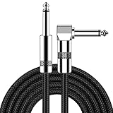
- [Low Noise High Fidelity] New bee guitar cord with 22AWG oxygen-free-copper center conductors provides enhanced signal transfer, the PVC insulation material and the high-density stranded copper double shield provide noise rejection for quiet operation
- [Ideal for Choice] New bee 1/4 straight and right angle metal connectors, ensures a secure connection in any instrument, pedal, or amp jack. L shape great fro on stage guitar stands
- [Wide Compatible] Suitable for mixer, power amplifier, microphone, effector, decoder, equalizer, electoronic organ, electric guitar, electric bass and other electronic equipments. Widely used in the installation of smart home, karaoke speaker, professional stage audio, etc
- [Easy Disassemble] Making incidental repairs quick and easy! Very nice, flexible cable
- [Dont Hesite to Order] 24 hours customer services and professional technology team are standing by
When MRI and PET appear to point in different directions, that disagreement is a reason for careful review rather than a reason to assume that one modality always wins. The team may review image quality, protocol details, prior studies and clinical timing, then decide whether additional follow-up or another evaluation is needed. The cited guidance supports using the modalities for their complementary information; it does not provide a universal rule for resolving every discordant result.
Recommended Free Tools
Common misunderstandings about MRI versus PET
“PET is more advanced, so it should replace MRI.”
The cited glioma guideline says the opposite of a blanket replacement model: MRI is the primary clinical imaging modality across glioma disease stages. PET can supplement MRI for selected questions, particularly when conventional MRI does not clearly distinguish recurrence from treatment-related change.
“Any PET scan provides the same answer.”
PET findings depend on the tracer and protocol. Amino-acid PET and FDG PET are not presented as identical tools, and the brain-metastasis guidance gives amino-acid PET a more prominent role for the recurrence-versus-treatment-effect question.
“A trial schedule is the standard schedule for everyone.”
PET RANO 1.0 is a proposed framework for diffuse-glioma clinical trials. Its intervals and paired PET/MRI approach should not be converted into a personal schedule without direction from the treating team.
“One scan can determine recurrence by itself.”
Imaging is interpreted with treatment timing, prior examinations and clinical findings. PET may add information, but the scan is not a stand-alone verdict.
Do these 3 things before closing this tab:
1Repair Windows errors before they cause bigger problems2Scan for outdated or missing drivers - takes under a minute3Clear out junk files and repair common Windows errors“Guidance for glioma automatically applies to every brain tumor.”
The strongest guidance summarized here concerns gliomas and brain metastases. It does not establish one approach for every primary or secondary brain tumor.
What determines an individual monitoring plan
There is no single MRI-versus-PET schedule established for all brain tumors. The strongest guidance summarized here concerns gliomas and brain metastases; it does not establish one approach for every primary or secondary brain tumor. A neuro-oncology or oncology team selects imaging in the context of the tumor type, treatment, clinical findings and prior imaging, and the question that needs an answer.
- Tumor type: The cited guidance treats glioma and brain metastases in distinct recommendations; findings for one should not be assumed to apply identically to every tumor.
- Treatment and timing: The glioma guideline includes MRI in settings ranging from early postsurgical assessment to surveillance during chemotherapy. The relevant clinical stage affects what the imaging is meant to assess.
- Clinical findings: Imaging is interpreted with the clinical context, not as a substitute for it.
- Prior scans: The team considers changes over time under the response framework relevant to the tumor and modality.
- The unresolved question: If the concern is structural change, MRI is central in the cited glioma guidance. If MRI leaves a recurrence-versus-treatment-effect question unclear, selected PET use may add information.
- Local availability and protocol: PET tracer and protocol matter, and what can be used depends in part on local availability.
These factors are reasons to discuss the rationale for a scan with the treating team, not a basis for requesting or rejecting PET or MRI on the comparison alone. The EANO guideline index provides a route to additional disease-specific guidance.
Independent reader supportYour contribution helps us test, update, and keep practical guides available for everyone.Questions to ask the care team about a scan
If the purpose of a proposed scan is unclear, focused questions can help connect the test to the decision it is meant to inform:
Free tools Windows power users keep installed
One-click scans. No signup required.
Rank #4

- Digital Family Calendar: BSIMB digital calendar wall touch screen puts an end to your chaotic life. Designed for busy families, the digital calendar keeps you organised and in step with your family. Assign different colours and tasks to each family member to make family scheduling easier and more intuitive.
- Easy Setup and Auto-Sync: Plug in, connect to WLAN and auto-syn your electric calendar in minutes. Digital wall calendar compatible with Google calendar, iCloud calendar, Outlook calendar, Cozi calendar and Yahoo calendar. Add events directly on the device or through the ecalendar app. Electronic calendar stays current whether you're at work or traveling—no manual updates needed.
- All-In-One Home Assistant: Smart calendar builds habits and encourage independence with personalised chores. Display daily meal plan to reduce the daily ‘What's for dinner?’ stress. To-do lists keep track of everything you want. Turning chores into valuable achievements with stars and rewards, making life easier and more enjoyable.
- Manage Anytime Anywhere: BSIMB electronic calendar for family stay connected with the powerful ecalendar app. Access and manage your digital wall calendar anytime, anywhere with free features like stars & rewards, photo screensaver, chore charts, meals plans, to-do lists etc. Electronic planner paid features like Magic Import tailor advanced tools for users with special needs.
- Stay in Touch with Loved Ones: Share photos or videos from your phone to electronic wall calendar with app, instantly update your life moment, and keep in touch with your family, especially those who are far away; you can also surprise your smart calendar digital wall by sending him a wish card; it's also great for keeping tech-illiterate elders in touch with the youngsters.
- What specific question is this scan intended to answer?
- Is the main concern structural change, possible recurrence, treatment effect or response to therapy?
- How will the result be interpreted alongside prior imaging and clinical findings?
- If PET is proposed, which tracer and protocol are being used, and what information might they add to MRI in this case?
- Is the schedule being discussed part of a clinical-trial protocol or the person’s routine care?
- Will the current examination be compared with a defined baseline or with several prior scans?
- What would a clear, unclear or conflicting result mean for the next decision?
- What will the team consider if the imaging remains inconclusive?
These questions do not tell a person which scan to choose. They help clarify the clinical purpose, the limits of the result and how the team plans to use it.
Frequently asked questions
Which is better for monitoring a brain tumor, MRI or PET?
There is no single answer for every tumor. MRI is generally the primary monitoring modality for glioma and is used across evaluation, treatment planning, surveillance and recurrence assessment. PET can add metabolic information for selected questions, so it is usually complementary rather than universally better or a replacement for MRI.
When is a PET scan used instead of an MRI?
The cited guidance does not establish PET as a universal alternative to MRI. Amino-acid PET is described as an addition to MRI when conventional MRI makes it difficult to distinguish recurrent brain metastasis from treatment-related change. The treating team decides whether that additional information is relevant.
What is the difference between amino-acid PET and FDG PET in this context?
The glioma guidance discusses MET, FET and FDOPA, which are radiolabeled amino-acid tracers, as well as FDG. For the specific recurrence-versus-treatment-effect problem in brain metastases, the 2025 guidance gives amino-acid PET a more prominent role than FDG PET. Tracer choice remains protocol- and question-dependent.
Windows Errors? Fix Them Before They Spread
Repair common Windows errors and clear accumulated junk for a smoother, more stable PC - no reinstall needed.Free scan · no reinstallCrashes, No Sound, or Screen Glitches?
Random freezes, missing sound and display glitches usually trace back to one bad driver. Find and replace yours safely.Free scan · under a minuteDoes a PET scan prove that a tumor has returned?
No. PET may contribute metabolic evidence, but interpretation also considers MRI, prior images, treatment timing and clinical findings. A result should be discussed with the treating team rather than treated as a stand-alone verdict.
Does every person with a brain metastasis need PET?
No. The brain-metastasis guideline describes amino-acid PET as useful in addition to MRI for selected cases in which recurrence and treatment-related change are difficult to separate. It does not recommend that every person undergo PET.
Are PET RANO 1.0 intervals routine follow-up rules?
No. PET RANO 1.0 is a proposed amino-acid PET response framework for diffuse-glioma clinical trials. Its proposed intervals vary by tumor grade and should not be presented as a universal routine-care schedule.
Why might two patients receive different scans?
Monitoring depends on tumor type, treatment, clinical findings, prior imaging, the unresolved question and local tracer or protocol availability. Guidance for glioma and brain metastases is not automatically applicable to every other brain tumor.
What should I do if MRI and PET reports seem to conflict?
Ask the treating team to review both reports and the actual images in context. The cited guidance supports complementary use of structural and metabolic information but does not prescribe one rule for every discordant result.
Limits of the comparison
The cited guidelines support a clear overall distinction—MRI is primary for glioma monitoring, while PET can supplement MRI for selected questions—but they do not provide a universal scan schedule, a single best tracer for every patient, or a rule for deciding an individual’s care. The brain-metastasis guidance addresses a particular challenge, and PET RANO 1.0 is a proposed clinical-trial response framework for diffuse glioma. Individual interpretation belongs to the treating team, which can account for the tumor, treatment, imaging history and clinical picture.
No comparison table can replace review of the actual images and the clinical record. The most useful way to read a recommendation is to ask what problem it addresses, which tumor population it concerns, which tracer or protocol it assumes and whether it applies to routine care or a trial. Those distinctions keep MRI’s established monitoring role clear while showing where PET may provide meaningful additional information.
Frequently Asked Questions
Which is better for monitoring a brain tumor, MRI or PET?
MRI is generally the primary monitoring test for glioma. PET can add metabolic information for selected questions, so it is usually complementary rather than a universal replacement.
When is PET used in addition to MRI?
Amino-acid PET may be added when conventional MRI does not clearly distinguish recurrent brain metastasis from treatment-related change, subject to the treating team’s judgment.
Does PET RANO 1.0 set everyone’s follow-up schedule?
No. It is a proposed response framework for diffuse-glioma clinical trials, not a universal routine-care timetable.
Does every PET scan use the same tracer?
No. Guidance discusses amino-acid tracers such as MET, FET and FDOPA as well as FDG, and the appropriate choice depends on the clinical question and local protocol.
Can one scan prove recurrence?
No. MRI or PET findings must be interpreted with treatment timing, prior images and clinical findings.
Free tools Windows power users keep installed
One-click scans. No signup required.
The Bottom Line
Bottom line: MRI is generally the backbone of brain-tumor monitoring, particularly for glioma. PET—especially amino-acid PET—may add useful metabolic information when MRI leaves questions such as recurrence versus treatment effect unresolved. Tracer, protocol, tumor type, treatment and clinical context all matter, so the treating team should determine whether PET adds value in an individual case.
Quick Recap
Product prices and availability are accurate as of the date/time indicated and are subject to change. Any price and availability information displayed on Amazon at the time of purchase will apply.

