Microscopy images can help identify what genes do when every image is tied to a known genetic perturbation. A practical workflow combines CRISPR perturbations, multiplexed fluorescence imaging, image preprocessing, machine-learning representations and comparisons with controls. The result is a ranked set of candidate functions, pathways and gene relationships—not proof of a complete molecular mechanism.
This guide explains how to design that workflow, what the leading examples actually measured, how to interpret model outputs, and which checks are needed before treating an image-based prediction as biology.

As an Amazon Associate I earn from qualifying purchases.
What image-based gene-function analysis can tell you
A cell’s shape, organelle distribution, texture and intensity can change after a gene is disrupted. If those changes are measured consistently across many cells, they form a phenotype that can be compared with phenotypes produced by other perturbations. Genes whose perturbations produce similar profiles may participate in the same pathway, complex or cellular process.
Outdated Drivers Are Slowing You Down
One free scan finds every outdated or missing driver and matches the right update for your exact hardware.Free scan · exact hardware matchWindows Errors? Fix Them Before They Spread
Repair common Windows errors and clear accumulated junk for a smoother, more stable PC - no reinstall needed.Free scan · no reinstallThat inference is conditional. A phenotype is visible only in the chosen cell type, culture condition, perturbation strength, time point and imaging panel. A gene can have an important function that is redundant, too subtle for the assay or visible only under stress. Morphological similarity therefore supports a related observable phenotype; it does not by itself prove direct physical interaction, causality between two genes or the complete mechanism of either gene.
#1 Best Overall

- [Ultra-Lightweight Ear Buds Designed for Small Ears] Each earbud weighs only 3.7g and features a compact, ergonomic in-ear design made especially for small ears. Secure, low-profile, and comfortable for workouts, all-day wear or overnight sleeping.The ultra-slim profile sits flush in your ear with no hard edges poking or rubbing, so you can even sleep on your side without discomfort.
- [Immersive Stereo Sound with TOZO OrigX Technology] TOZO OrigX tuning delivers clear vocals, balanced mids, and natural stereo sound for music, podcasts, and videos.
- [Long Battery Life for Daily Use] Get up to 7 hours of playtime on a single charge, with up to 32 hours total using the charging case—ideal for workdays, commuting, and extended listening sessions.
- [Bluetooth 5.3 & Stable Connection] Bluetooth 5.3 provides fast pairing, stable wireless performance, and reduced dropouts as you move around home or office.
- [Deep Bass with Clear Vocals] High-performance drivers produce punchy bass while keeping vocals clean and detailed for everyday listening.
The end-to-end workflow
1. Choose genes, perturbations and an assay context
Start by defining the biological question and the context in which a function should be visible. CRISPR guide RNAs can disable selected genes, activate them or introduce other perturbations, but the choice affects the phenotype. Knockout, knockdown and drug treatment differ in timing, residual activity and off-target risk. Record the cell type, passage range, growth conditions, perturbation duration and imaging time point as part of the experiment, not as metadata added later.
Include non-targeting guides, untreated or vehicle controls and, where possible, positive controls with established effects. Use multiple guides per gene and biological replicates on different plates or days. These controls let you distinguish a gene-specific morphology from plate position, batch, toxicity or editing-efficiency effects.
2. Link each cell to its genetic perturbation
In a pooled optical screen, many guide RNAs are introduced into one cell population. The guide identity must then be read by a barcode or in situ sequencing so that the phenotype of an individual cell can be associated with the gene it received. This identity link is the essential bridge between an image and a gene-function hypothesis; without it, a beautiful image is only an unlabeled observation.
The PERISCOPE description from the Broad Institute of MIT and Harvard reports guide RNAs targeting about 20,000 genes. Guide-derived barcodes identify the disabled gene in each cell, allowing a genome-scale morphology atlas. Pooled designs increase scale, but they also introduce incomplete editing, mixed guide identities and label noise. Measure guide representation and editing or knockdown performance when possible, and flag cells with ambiguous or missing labels rather than forcing them into a gene group.
3. Capture many cellular structures with Cell Painting
Cell Painting uses a set of fluorescent stains that mark multiple compartments, such as nuclei, cytoplasm, mitochondria, endoplasmic reticulum and other structures. Multiplexing records shape, texture, intensity and spatial relationships across the cell instead of relying on one pathway-specific reporter. A standard widefield microscope can be sufficient for the described PERISCOPE workflow, which used five Cell Painting stains together with four-color barcodes.
Rank #2

- 【IP68 Waterproof Multifunctinal Smart Watch】Torjalph fitness activity tracker smart watch is IP68 waterproof technology ,you can wear it when swimming,handwashing,it's rain-proof which means that you perform multiple exercise modes outside without worrying about sweat or rain corroding your fitness smart watch,and this watch equipped with a 230 mAh large capacity battery magnetic charging ,and this sports activity tracker watch can be used 5-7 days If fully charged.
- 【Larger 1.83'' Full Screen Watch & Multi-dial Selection】1.83 inch full HD color touch screen smart watch for men / women provides excellent 240*284 resolution image quality ,This Bluetooth smart watch offers a better touch experience and remarkable immersion and ,more than 200 watch faces for you to choose from App"Da Fit ", you can change the dial style according to your mood to wear keeping the world on your wrist
- 【Accurate Activity Tracker Smart Watch 】sports watch activity tracker uses sweat and dust resistant breathable silicone strap, our Fitness Tracker Smart Watch for Android iOS Phones supports 12 sports modes,walking,running,bicycle,swimming,badminton,basketball,football,rope skipping. This bluetooth calls smart watch for men women is lightweight and stylish, bringing more convenient and relaxing sports experience while also providing more protection.
- 【Your 24 Hour Personal Daily Health Monitor】Fitness watch for women men full touch screen IP67 waterproof bluetooth smartwatch for Android iOS Phones with heart rate blood pressure monitor multifunction Andriod smart watch with steps counter sleep monitor sports fitness watch, monitoring blood oxygen saturation allows you to understand your physical condition in real time to track fitness progress ,to know your health status in time.
- 【More Functions Bluetooth Call Smart Watch】This fitness activity smart watch full screen for iPhone and Android phones includes a bunch of new features including:The bluetooth smart watch can hang up the phone, receive and display WhatsAPP /Text /Facebook /Twitter /Messenger reminder.Support sedentary reminder, alarm clock, stopwatch, timer,weather information,music control, remote camera control compatible with Android 5.0 and above or iOS 9.0 and above smartphones.
Staining and sequencing order may need to be adapted in a pooled optical screen. The CellPaint-POSH study changed staining and sequencing components to preserve RNA signals while retaining image quality. Treat the exact stain panel, microscope settings, exposure limits and sequencing chemistry as part of the assay definition: changing them can change which gene effects are detectable.
4. Acquire images with controls for optical variation
Capture enough fields and cells per perturbation to estimate within- and between-plate variation. Keep focus, illumination, magnification, exposure and channel order consistent. Randomize perturbations across plate positions when practical, and distribute controls across the plate rather than placing them in one row or column.
The Tool Desk
Outbyte Driver Updater FREEFix the driver behind crashes, sound loss and screen glitchesFind Drivers →Outbyte PC Repair FREERepair Windows errors before they cause bigger problemsFix Now →Save raw images and acquisition metadata. Do not overwrite the original files with corrected images. A later model may need a different correction, and retaining raw data makes failed preprocessing recoverable.
5. Correct images and segment cells
Uneven illumination can make identical cells appear different in different parts of a field. CellPaint-POSH describes illumination correction followed by percentile normalization. Segmentation with CellPose identifies cell and nuclear boundaries, after which the workflow creates cropped single-cell image tiles. State these choices when reporting a result because preprocessing changes the measured phenotype.
Inspect segmentation visually on every plate and quantify failure rates. Common problems include merged adjacent cells, a nucleus assigned to the wrong cytoplasm, truncated cells at image borders and debris mistaken for cells. Exclude or separately label failed objects; silently retaining them can create a model that learns segmentation artifacts rather than biology.
Rank #3
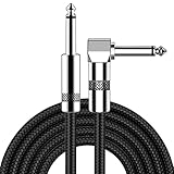
- [Low Noise High Fidelity] New bee guitar cord with 22AWG oxygen-free-copper center conductors provides enhanced signal transfer, the PVC insulation material and the high-density stranded copper double shield provide noise rejection for quiet operation
- [Ideal for Choice] New bee 1/4 straight and right angle metal connectors, ensures a secure connection in any instrument, pedal, or amp jack. L shape great fro on stage guitar stands
- [Wide Compatible] Suitable for mixer, power amplifier, microphone, effector, decoder, equalizer, electoronic organ, electric guitar, electric bass and other electronic equipments. Widely used in the installation of smart home, karaoke speaker, professional stage audio, etc
- [Easy Disassemble] Making incidental repairs quick and easy! Very nice, flexible cable
- [Dont Hesite to Order] 24 hours customer services and professional technology team are standing by
6. Represent each cell or perturbation as data
You can describe images with engineered measurements—area, intensity, texture, shape and compartment relationships—or with learned embeddings produced by a neural network. Aggregate single-cell measurements carefully. Median or robust mean profiles are less sensitive to a few damaged cells than an ordinary mean, while distributions can preserve biologically useful heterogeneity.
CellPaint-POSH compared classical morphological features, an ImageNet-trained DINO model and self-supervised DINO vision-transformer models trained on Cell Painting images. Its Cell Painting-trained representation performed better on the reported classification and gene-association tasks than the comparison representations. The lesson is not that one architecture always wins; it is that representation training data should resemble the assay and the biological variation the model must recognize.
7. Compare perturbation phenotypes
Normalize profiles using controls from the same batch, then calculate distances or similarities between perturbations. Cluster genes with similar profiles and test whether clusters are enriched for known pathways or complexes. A useful analysis reports the similarity metric, normalization, number of cells retained, replicate handling and how significance or cluster stability was assessed.
Similarity can recover expected relationships and reveal candidates. CellPaint-POSH reported clusters involving mitochondrial translation, cytoskeletal organization, lipid metabolism and mTORC1 signaling, as well as recovery of known gene associations. These are starting points for experiments: a cluster may reflect a shared pathway, a common stress response or a technical confounder.
8. Validate a candidate function
Validation should use an assay that measures the proposed function rather than repeating the same image analysis. For a mitochondrial candidate, measure respiration or mitochondrial translation; for a cytoskeletal candidate, test organization or dynamics with an independent assay; for a signaling candidate, measure pathway activity or response to a selective perturbation.
Free tools Windows power users keep installed
One-click scans. No signup required.
Rank #4

- Digital Family Calendar: BSIMB digital calendar wall touch screen puts an end to your chaotic life. Designed for busy families, the digital calendar keeps you organised and in step with your family. Assign different colours and tasks to each family member to make family scheduling easier and more intuitive.
- Easy Setup and Auto-Sync: Plug in, connect to WLAN and auto-syn your electric calendar in minutes. Digital wall calendar compatible with Google calendar, iCloud calendar, Outlook calendar, Cozi calendar and Yahoo calendar. Add events directly on the device or through the ecalendar app. Electronic calendar stays current whether you're at work or traveling—no manual updates needed.
- All-In-One Home Assistant: Smart calendar builds habits and encourage independence with personalised chores. Display daily meal plan to reduce the daily ‘What's for dinner?’ stress. To-do lists keep track of everything you want. Turning chores into valuable achievements with stars and rewards, making life easier and more enjoyable.
- Manage Anytime Anywhere: BSIMB electronic calendar for family stay connected with the powerful ecalendar app. Access and manage your digital wall calendar anytime, anywhere with free features like stars & rewards, photo screensaver, chore charts, meals plans, to-do lists etc. Electronic planner paid features like Magic Import tailor advanced tools for users with special needs.
- Stay in Touch with Loved Ones: Share photos or videos from your phone to electronic wall calendar with app, instantly update your life moment, and keep in touch with your family, especially those who are far away; you can also surprise your smart calendar digital wall by sending him a wish card; it's also great for keeping tech-illiterate elders in touch with the youngsters.
Reperturb the gene with an independent guide or reagent, rescue the phenotype with a guide-resistant construct when feasible, and test more than one cell background or condition if the claim is intended to generalize. Confirm that the effect is not explained by cell death, cell-cycle composition, guide toxicity or altered cell density.
Three approaches and what their numbers mean
| Approach | Biological input and labeling | Model or analysis | Reported result and scope |
|---|---|---|---|
| PERISCOPE genome-scale optical screening | Pooled CRISPR guides, guide-derived barcodes, five Cell Painting stains and four-color barcode imaging on a described widefield workflow. | Image-based profiling of cellular morphology linked to guide identities. | Broad Institute reporting describes more than 20,000 genes and hundreds of image-based cellular features. It also reports data generation as more than ten times less expensive than comparable high-dimensional approaches such as high-throughput single-cell RNA sequencing; that is a comparison under the report’s costing assumptions, not a universal laboratory price. |
| CellPaint-POSH | Pooled Cell Painting CRISPR screening with assay-specific staining and sequencing changes to preserve RNA signals. | Classical features, ImageNet-trained DINO and Cell Painting-trained self-supervised DINO vision-transformer embeddings; similarity and clustering for gene relationships. | The study’s methods describe approximately 1.585 million cells for CP-DINO-300 and 1.559 million for CP-DINO-1640. Cell Painting-trained embeddings performed better on the reported tasks and recovered known associations; these are study-specific evaluations. |
| Image2Reg | Chromatin images paired with molecular information about gene interactions; this is a separate design from pooled Cell Painting. | Image and gene representations are aligned so the model can infer perturbations, including perturbations absent from training. | A Broad Institute report describes 60% accuracy for predicting genetic drug targets, including compounds not seen during training. The evaluation used one cell type and specific perturbation conditions, so it is not a general microscopy-AI benchmark. |
These approaches should not be treated as interchangeable. PERISCOPE and CellPaint-POSH associate multiplexed cell morphology with pooled perturbation identities. Image2Reg aligns chromatin images with molecular gene information and addresses a different prediction problem. Compare the biological input, labeling method, cell type, perturbation design, training data and validation split before comparing percentages.
Independent reader supportYour contribution helps us test, update, and keep practical guides available for everyone.Designing trustworthy controls and quality checks
Controls for biology
- Non-targeting guides: estimate the baseline distribution and detect guide-independent effects.
- Known positive perturbations: test whether the panel can recover an expected morphology.
- Multiple guides and replicates: separate gene effects from guide-specific or day-specific effects.
- Rescue or orthogonal perturbation: test whether restoring or independently changing the gene changes the phenotype as predicted.
Controls for imaging and computation
- Distribute controls across plates and positions to expose illumination, edge and batch effects.
- Track focus, saturation, missing channels, staining intensity and segmentation quality.
- Report how many cells and perturbations were removed and why.
- Keep training and test data separated by biological replicate or batch when measuring generalization; random cell-level splits can leak nearly identical conditions into both sets.
- Check whether a classifier is learning cell number, debris, plate position or toxicity instead of the intended phenotype.
How to interpret an AI prediction
Read a prediction as a ranked hypothesis. Ask which cells contributed to it, how consistent the phenotype was across guides and replicates, which controls define the baseline, and whether the model’s nearest neighbors are biologically plausible. Examine representative images from both high-similarity and low-similarity groups. A cluster that disappears after batch correction is a technical result, not a gene relationship.
Negative results require equal care. “No detectable phenotype” means no reliable difference under this assay’s sensitivity and context. It does not establish that the gene is inactive or unimportant. Conversely, a strong phenotype can reflect general toxicity or cell-cycle arrest rather than the gene’s specific function. Report effect size, uncertainty and the tested context rather than only a top-hit list.
Do these 3 things before closing this tab:
1Repair Windows errors before they cause bigger problems2Fix the driver behind crashes, sound loss and screen glitches3Clear out junk files and repair common Windows errorsPractical implementation checklist
- Define the cell type, perturbation modality, time point and biological question.
- Select guides, non-targeting controls, positive controls and replicate structure.
- Plan how guide or barcode identities will be assigned to individual cells.
- Choose a multiplexed stain panel and lock microscope acquisition settings.
- Acquire raw images with plate-position randomization and complete metadata.
- Apply documented illumination correction and normalization; retain raw files.
- Segment nuclei and cells, inspect failures and generate single-cell tiles or feature tables.
- Build a baseline with classical features before or alongside learned embeddings.
- Train or select representations using biologically appropriate data, avoiding leakage between train and test replicates.
- Aggregate cells by perturbation, calculate similarities and test cluster stability.
- Compare candidates with known gene associations and pathway annotations.
- Validate leading hypotheses with an independent molecular or functional assay.
Common failure modes and fixes
The model separates plates instead of genes
Plate-specific illumination, staining or density is often the cause. Improve randomization, apply correction using appropriate controls, include batch-aware validation and inspect embeddings colored by plate before interpreting gene clusters.
Guide identities are unreliable
Low barcode signal, incomplete editing and multiple guides per cell can create label noise. Set identity-quality thresholds, report the fraction of assigned cells, use guide-level concordance and analyze ambiguous cells separately.
Segmentation creates false phenotypes
Over- or under-segmentation changes area, shape and intensity features. Tune CellPose or another segmenter on representative images, quantify errors by condition and exclude fields with systematic failures.
A model performs well but does not generalize
A random split can make the task easier than a new experiment because cells from the same plate share artifacts. Test on held-out biological replicates, plates or perturbations. For Image2Reg-style claims about unseen perturbations, preserve the exact evaluation definition and cell context used in the report.
Quick wins for a faster PC:
Fix the driver behind crashes, sound loss and screen glitchesFind Drivers →Clear out junk files and repair common Windows errorsFree Scan →A similar phenotype is mistaken for a direct interaction
Shared stress, toxicity or convergent downstream effects can make unrelated genes look similar. Use orthogonal assays, rescue experiments and perturbation combinations before claiming a direct relationship.
What the published examples support—and what they do not
The cited studies show that high-content morphology can be scaled, linked to genetic perturbations and analyzed with learned representations to recover known associations and propose new ones. PERISCOPE’s reported scale, CellPaint-POSH’s large training sets and Image2Reg’s reported 60% target-prediction accuracy demonstrate specific capabilities under defined conditions.
They do not establish a universal cost, accuracy or transferability for every microscope, stain panel, cell type or perturbation library. Image2Reg was evaluated in one cell type and specified perturbation conditions, and adaptation to other contexts remains prospective. CellPaint-POSH’s results likewise depend on its image processing, training data, controls and assay design.
Bottom line for a new project
Use microscopy and AI when the function you seek is likely to produce a measurable cellular phenotype and you can maintain a reliable perturbation-to-cell identity link. Build the experiment around controls, replicates, documented preprocessing and an independent validation assay. Train representations on data that match your imaging biology, treat similarity as a hypothesis generator, and carry the cell type, perturbation and evaluation context with every reported number. That combination turns images into evidence about gene function without overstating what morphology alone can prove.
Quick Recap
Product prices and availability are accurate as of the date/time indicated and are subject to change. Any price and availability information displayed on Amazon at the time of purchase will apply.

