Read a single-cell embryo atlas as a reference of sampled cells and measured molecular states—not as a complete census or a literal map of embryonic fate. Start by checking the species, staging convention, tissue coverage, embryos sampled, assay and time points. Then determine how cell labels were assigned, what the two-dimensional map actually encodes, whether spatial context was preserved, and whether any lineage path is inferred from expression or measured by ancestry tracing. Those checks prevent the most common errors: treating human and mouse stage labels as interchangeable, assuming a marker proves a cell identity, or reading a visual branch as proof that one cell became another.
What a single-cell embryo atlas actually contains
A single-cell atlas is built from cells (or, in some studies, nuclei) collected from embryos and profiled for molecular features, usually transcript abundance. Computational analysis groups cells with similar measured profiles, assigns labels to those groups, and displays the relationships in a chart. The result is a structured sample of molecular states observed under a particular experiment’s collection, dissociation, sequencing and analysis choices.

As an Amazon Associate I earn from qualifying purchases.
That distinction matters. An atlas is not a complete inventory of every cell in every embryo, and a dot on a plot is not a physical coordinate in the embryo. A cluster may be missing because a tissue was not sampled, a cell was lost during preparation, or the method captured it poorly. A state may appear unusually large because it was easier to recover or because the study sampled a time when it was abundant.
Most atlas pages combine several layers:
- Measurements: a matrix of features for each captured cell or nucleus.
- Groups: clusters or other partitions based on similarity in those measurements.
- Annotations: names assigned to groups or individual cells, often at several levels of detail.
- Embeddings: two-dimensional layouts such as UMAP, t-SNE or force-directed plots that make high-dimensional similarity visible.
- Developmental context: stage, collection time, embryo identity and sometimes anatomical location.
- Lineage analysis: a proposed progression from expression and time, or direct ancestry information from a tracing experiment.
Interpret each layer separately before combining them. A label is an interpretation of measurements; an embedding is a visualization of similarity; a trajectory is a model of relationships. None should be silently upgraded into a direct observation of anatomy or fate.
#1 Best Overall

- [Ultra-Lightweight Ear Buds Designed for Small Ears] Each earbud weighs only 3.7g and features a compact, ergonomic in-ear design made especially for small ears. Secure, low-profile, and comfortable for workouts, all-day wear or overnight sleeping.The ultra-slim profile sits flush in your ear with no hard edges poking or rubbing, so you can even sleep on your side without discomfort.
- [Immersive Stereo Sound with TOZO OrigX Technology] TOZO OrigX tuning delivers clear vocals, balanced mids, and natural stereo sound for music, podcasts, and videos.
- [Long Battery Life for Daily Use] Get up to 7 hours of playtime on a single charge, with up to 32 hours total using the charging case—ideal for workdays, commuting, and extended listening sessions.
- [Bluetooth 5.3 & Stable Connection] Bluetooth 5.3 provides fast pairing, stable wireless performance, and reduced dropouts as you move around home or office.
- [Deep Bass with Clear Vocals] High-performance drivers produce punchy bass while keeping vocals clean and detailed for everyday listening.
First establish the atlas’s scope
Before reading a cluster name, identify exactly what the dataset sampled. Record the organism, embryo or tissue fraction, developmental range, number of embryos, sampling interval and whether extra-embryonic tissues or later body parts were included. These details determine what conclusions are possible.
Species and staging convention
Developmental labels are species-specific. “E7.5” and “E8.5” are mouse embryonic-day notation; they are not interchangeable with a human embryonic day. Human early-organogenesis datasets may use Carnegie stages, which are morphological stages rather than a simple day count. Translate neither system by number alone. Use the staging convention stated by the study and, when comparing studies, compare biological landmarks and sampled anatomy as well as nominal labels.
Embryos, tissues and sampling intervals
Ask whether the atlas used whole embryos, selected regions, extra-embryonic tissues, organ-specific material or a mixture. A whole-embryo atlas can show relationships among systems, while a dissected-tissue atlas can provide finer resolution within one region but cannot support claims about unsampled tissues. Also check whether time points are evenly spaced. A gap between collections can hide a short-lived state; a half-day series can reveal changes that a two-day series misses.
Dataset examples and why their counts are not rankings
| Study or resource | Organism and window | Reported scale | What the figure does—and does not—tell you |
|---|---|---|---|
| MELA project page (2026) | Mouse embryos, E7.5–E10.0, half-day stages | More than 1.5 million cells from 16 embryos; roughly 75% of cell divisions resolved | Reconstructed lineage trees are paired with deep transcriptional profiling in this mouse experiment. The scale and resolution apply to that project, not to mouse atlases generally. |
| Xu et al. (2023), NCBI BioProject PRJNA660958 | Human early organogenesis, Carnegie stages 12–16 | 185,140 single-cell transcriptomes; 313 cell types across 18 developmental systems | The record describes a particular human window and an annotation file updated December 5, 2022. It is not a count for all human embryo atlases. |
| Pijuan-Sala et al., Nature (2019) | Mouse, nine time points from E6.5 to E8.5 | 116,312 cells | The study used temporal and transcriptional information to build a molecular map toward major embryonic lineages. |
| Cao et al., Nature Cell Biology (2026), mdCLA | Mouse, E1.5–E19.0, 37 time points | 2,230,900 cells after quality control from 351 embryos; six broad groups, 108 organs or tissues, 305 cell types and 1,316 subtypes | The hierarchy and marker list describe this atlas’s scope and annotation choices. Its larger cell count does not by itself make it better for every question. |
Cell totals are affected by embryo number, stage range, capture efficiency, sequencing depth, pooling and whether the study profiles cells or nuclei. Compare those design choices before treating one atlas as more comprehensive or more precise.
How to read developmental stages without mixing species
Find the study’s staging key, not just the label shown beside a cluster. For each time point, note the organism, the convention, the anatomical or morphological criteria used and the actual collection range. In a human dataset covering Carnegie stages 12–16, a stage number refers to a defined morphological window. In a mouse series labeled E6.5–E8.5, the number refers to embryonic day under a mouse-specific convention. Similar-looking numbers do not imply equivalent developmental events.
Rank #2

- 【IP68 Waterproof Multifunctinal Smart Watch】Torjalph fitness activity tracker smart watch is IP68 waterproof technology ,you can wear it when swimming,handwashing,it's rain-proof which means that you perform multiple exercise modes outside without worrying about sweat or rain corroding your fitness smart watch,and this watch equipped with a 230 mAh large capacity battery magnetic charging ,and this sports activity tracker watch can be used 5-7 days If fully charged.
- 【Larger 1.83'' Full Screen Watch & Multi-dial Selection】1.83 inch full HD color touch screen smart watch for men / women provides excellent 240*284 resolution image quality ,This Bluetooth smart watch offers a better touch experience and remarkable immersion and ,more than 200 watch faces for you to choose from App"Da Fit ", you can change the dial style according to your mood to wear keeping the world on your wrist
- 【Accurate Activity Tracker Smart Watch 】sports watch activity tracker uses sweat and dust resistant breathable silicone strap, our Fitness Tracker Smart Watch for Android iOS Phones supports 12 sports modes,walking,running,bicycle,swimming,badminton,basketball,football,rope skipping. This bluetooth calls smart watch for men women is lightweight and stylish, bringing more convenient and relaxing sports experience while also providing more protection.
- 【Your 24 Hour Personal Daily Health Monitor】Fitness watch for women men full touch screen IP67 waterproof bluetooth smartwatch for Android iOS Phones with heart rate blood pressure monitor multifunction Andriod smart watch with steps counter sleep monitor sports fitness watch, monitoring blood oxygen saturation allows you to understand your physical condition in real time to track fitness progress ,to know your health status in time.
- 【More Functions Bluetooth Call Smart Watch】This fitness activity smart watch full screen for iPhone and Android phones includes a bunch of new features including:The bluetooth smart watch can hang up the phone, receive and display WhatsAPP /Text /Facebook /Twitter /Messenger reminder.Support sedentary reminder, alarm clock, stopwatch, timer,weather information,music control, remote camera control compatible with Android 5.0 and above or iOS 9.0 and above smartphones.
Stage also describes the sampled embryo, not necessarily the age of every cell in it. Cells can retain earlier molecular programs, transition between states, or mature at different rates within the same embryo. A cluster that appears at one sampled stage may be genuinely transient, absent because of sampling, or difficult to capture. Look for the number of embryos per stage and whether the atlas shows biological replicates rather than a single pooled observation.
When a study spans many stages, examine both the broad stage panel and the per-stage cell counts. A late-stage increase in a cell type may reflect expansion, improved capture, inclusion of a tissue that was not present earlier, or a change from cell to nucleus preparation. The stage label is necessary context, but it is not an explanation by itself.
Recommended Free Tools
Decode the annotation hierarchy
Atlas labels usually form a hierarchy. A broad group might contain a tissue, which contains a cell type, which may contain several subtypes or states. “Neural,” “neural tube,” “neural progenitor” and a named neuronal subtype are different resolutions of description. Do not compare labels at different levels as if they were equivalent categories.
What supports a label
For every label, look for the evidence listed in the paper or atlas interface:
- Marker genes or a broader expression signature.
- The clustering or classification method used to separate the group.
- Whether experts curated the label manually, transferred it from a reference, or combined both approaches.
- Validation against another dataset, a spatial measurement or an established anatomical marker.
- The confidence or ambiguity assigned to borderline cells.
A marker is evidence, not a guarantee. A gene can be expressed in more than one lineage, switched on during a transition, or detected unevenly because of technical dropout. Conversely, a genuine cell may lack a canonical marker in a shallow measurement. Read a label as “cells whose measured profiles support this interpretation under the stated method,” not as an unmediated observation.
Rank #3
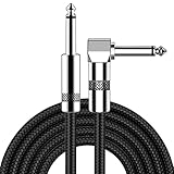
- [Low Noise High Fidelity] New bee guitar cord with 22AWG oxygen-free-copper center conductors provides enhanced signal transfer, the PVC insulation material and the high-density stranded copper double shield provide noise rejection for quiet operation
- [Ideal for Choice] New bee 1/4 straight and right angle metal connectors, ensures a secure connection in any instrument, pedal, or amp jack. L shape great fro on stage guitar stands
- [Wide Compatible] Suitable for mixer, power amplifier, microphone, effector, decoder, equalizer, electoronic organ, electric guitar, electric bass and other electronic equipments. Widely used in the installation of smart home, karaoke speaker, professional stage audio, etc
- [Easy Disassemble] Making incidental repairs quick and easy! Very nice, flexible cable
- [Dont Hesite to Order] 24 hours customer services and professional technology team are standing by
Manual and transferred annotations
The 2026 mdCLA paper describes manual annotation that combines transcriptomic signatures with literature markers in a four-level classification. That makes the hierarchy useful, but it also means the names embody expert decisions. A transferred label depends on the reference atlas and the similarity rule used to transfer it. If two studies use different references or different granularity, the same biological continuum can receive different names.
The Tool Desk
Outbyte Driver Updater FREEScan for outdated or missing drivers - takes under a minuteDriver Scan →Outbyte PC Repair FREEClear out junk files and repair common Windows errorsFree Scan →Atlas records can also change. The NCBI BioProject associated with Xu et al. records an annotation-file update dated December 5, 2022. When reproducing a result, save the version or date of the annotation file as well as the accession, because a later label revision can change counts or names without changing the underlying reads.
What clusters and maps mean
UMAP, t-SNE and force-directed layouts arrange cells according to a chosen representation of expression similarity. Nearby points generally have similar measured profiles under that analysis. The axes have no biological units, and the shape is not a tissue cross-section.
What you may infer
- Which cells have similar expression profiles in the displayed feature space.
- Whether a label is internally heterogeneous or contains several visible subgroups.
- Whether cells from different stages or embryos overlap or occupy distinct regions.
- Whether a proposed subtype is supported by a reproducible separation across replicates.
What the map cannot prove on its own
- Physical distance in the embryo.
- Direct developmental ancestry.
- The order in which cells changed state.
- That a large visual gap represents a fixed biological boundary.
Embedding parameters, selected genes, normalization and the number of cells can change the appearance. A branch in two dimensions may be a projection of a curved continuum, and a compact island may contain multiple states that separate only at higher resolution. Inspect the underlying marker plots, replicate behavior and alternative views before making a biological claim.
Use time and space together
A time course adds an ordering signal that a single snapshot lacks. If a population increases, changes markers and gives way to another population across successive samples, those observations can support a developmental model. They still do not demonstrate that every cell in the earlier group became every cell in the later group.
Free tools Windows power users keep installed
One-click scans. No signup required.
Rank #4

- Digital Family Calendar: BSIMB digital calendar wall touch screen puts an end to your chaotic life. Designed for busy families, the digital calendar keeps you organised and in step with your family. Assign different colours and tasks to each family member to make family scheduling easier and more intuitive.
- Easy Setup and Auto-Sync: Plug in, connect to WLAN and auto-syn your electric calendar in minutes. Digital wall calendar compatible with Google calendar, iCloud calendar, Outlook calendar, Cozi calendar and Yahoo calendar. Add events directly on the device or through the ecalendar app. Electronic calendar stays current whether you're at work or traveling—no manual updates needed.
- All-In-One Home Assistant: Smart calendar builds habits and encourage independence with personalised chores. Display daily meal plan to reduce the daily ‘What's for dinner?’ stress. To-do lists keep track of everything you want. Turning chores into valuable achievements with stars and rewards, making life easier and more enjoyable.
- Manage Anytime Anywhere: BSIMB electronic calendar for family stay connected with the powerful ecalendar app. Access and manage your digital wall calendar anytime, anywhere with free features like stars & rewards, photo screensaver, chore charts, meals plans, to-do lists etc. Electronic planner paid features like Magic Import tailor advanced tools for users with special needs.
- Stay in Touch with Loved Ones: Share photos or videos from your phone to electronic wall calendar with app, instantly update your life moment, and keep in touch with your family, especially those who are far away; you can also surprise your smart calendar digital wall by sending him a wish card; it's also great for keeping tech-illiterate elders in touch with the youngsters.
Spatial measurements add a different kind of evidence: where a cell or expression pattern occurs in the embryo. Dissociation-based single-cell measurements generally lose that location. A spatial assay or mapping step can test whether a transcriptomic label appears in the anatomical region expected for it, and can reveal neighboring populations that look similar in expression but occupy different positions.
When only one stage is shown, treat absence as “not observed in this sample,” not automatically “not present in development.” When a cluster appears only at one stage, consider three explanations in parallel: a transient biological state, a sampling gap or tissue-composition difference, and a capture or quality-control effect. Replicate embryos and spatial confirmation help distinguish them.
Tell an inferred trajectory from a measured lineage
“Trajectory” is used for more than one kind of evidence. Before describing a path as a lineage, identify how it was constructed.
| Evidence type | What it supports | What it does not establish by itself |
|---|---|---|
| Expression-based trajectory | A computational ordering of cells whose measured profiles resemble a progression. | That cells physically traveled along the path, or that one specific cell produced the next state. |
| Time-ordered atlas | Changes in population composition and expression across sampled stages. | Direct ancestry between cells from different embryos or time points. |
| Spatial mapping | Anatomical location and neighborhood relationships for a molecular state. | Heritable descent or the timing of a transition. |
| Heritable lineage tracing | Ancestry relationships recorded through a mark passed to descendants in the experiment. | A universal fate rule outside the traced organism, stages, tissue and perturbation conditions. |
Pijuan-Sala and colleagues wrote that they “construct a molecular map of cellular differentiation from pluripotency towards all major embryonic lineages, and explore the complex events involved in the convergence of visceral and primitive streak-derived endoderm.” That wording describes a molecular map: it is powerful evidence about changing states and relationships, but it should not be paraphrased as direct ancestry unless a separate tracing experiment supports that claim.
Do these 3 things before closing this tab:
1Repair Windows errors before they cause bigger problems2Scan for outdated or missing drivers - takes under a minute3Clear out junk files and repair common Windows errorsThe MELA project page describes reconstructed mouse lineage trees paired with deep transcriptional profiling and reports that the combination quantifies fate biases, restriction timing, progenitor pool sizes and lineage relationships across replicate embryos. Its trees come from a particular mouse experiment and a specific lineage-tracing approach. Use them as stronger ancestry evidence within that scope, not as proof that every single-cell atlas directly traces lineage.
How to read a branch
- Check whether the branch was calculated from expression, time, known markers, a tracing barcode or a combination.
- Find the root and terminal states defined by the authors; a graph’s visual left-to-right direction is not automatically chronological.
- Look for replicate support and uncertainty along the path.
- Ask whether intermediate cells were actually sampled or merely interpolated by the algorithm.
- Use cautious language unless ancestry was measured: “consistent with,” “suggests a transition” or “is ordered toward,” rather than “became.”
Compare two embryo atlases on evidence, not cell count
When two resources disagree, compare their design before deciding that one is wrong. Use the following axes:
| Axis | Questions to ask | Why it changes interpretation |
|---|---|---|
| Organism and stage system | Which species and staging convention are used? Are the biological landmarks comparable? | Nominal stage numbers can refer to different developmental events. |
| Coverage | Which stages and tissues are sampled, at what intervals, and from how many embryos? | A missing tissue or a long time gap can look like a biological absence. |
| Sampling and assay | Whole embryo or selected parts? Cells or nuclei? Which sequencing and capture approach? | Preparation changes which populations are recovered and how completely. |
| Annotation | What label hierarchy is used, and which markers or validation support it? | Different granularity can make equivalent populations appear to have different names or counts. |
| Spatial context | Are cells dissociated, measured in place, or mapped back to anatomy? | Location can separate states that expression alone makes similar. |
| Lineage evidence | Is ancestry inferred from expression and time, or measured with heritable tracing? | These methods answer related but distinct questions about fate. |
Capture method is a real source of bias
The mdCLA paper compares droplet-based and nucleus-based approaches and reports differences in representation of some cell groups, including a higher neural-cell proportion at later stages in the cited nucleus-based comparison. Treat that as a method caveat. It does not show that one preparation is universally superior; the appropriate choice depends on stage, tissue, cell fragility and the question being asked.
A step-by-step workflow for reading any atlas
- Write a one-line scope statement. Record species, embryo or tissue, stage range, number of embryos, assay, and whether cells or nuclei were profiled.
- Translate the stage labels within their own system. Mark human Carnegie stages, mouse E-stages or another convention separately; do not create a cross-species day scale by arithmetic.
- Inspect the sampling table. Check embryos per stage, pooled versus individual samples, tissue exclusions and intervals between collections.
- Read the annotation legend before the plot. Identify broad groups, tissues, cell types and subtypes, then find the markers and validation attached to each level.
- Check annotation provenance. Note manual curation, reference transfer, automated classification and any version or update date.
- View the embedding as similarity. Read colors and labels, but do not interpret distance as physical location or a branch as ancestry.
- Overlay time and embryo identity. Determine whether a group is reproducible across embryos, restricted to one stage, or driven by one sample.
- Look for spatial evidence. If cells were dissociated, find out whether a separate spatial experiment or anatomical mapping confirms their location.
- Classify the lineage claim. Label each path as expression-inferred, time-supported, spatially supported or directly traced.
- State the conclusion at the same resolution as the evidence. Name the population and evidence type, include the species and stage, and preserve uncertainty instead of converting a model into a fact.
Common interpretation failures and their fixes
| Failure | Why it fails | Better practice |
|---|---|---|
| “E8.5 is the same developmental age as human day 8.5.” | Stage systems and developmental rates are species-specific. | Use the study’s own convention and compare landmarks, not numbers. |
| “This marker proves the cell type.” | Markers can overlap, be transient or be missed by technical dropout. | Use marker combinations, cluster context and validation. |
| “The nearest cluster is the parent.” | Embedding proximity represents similarity under an analysis, not ancestry. | Check trajectory method and tracing evidence. |
| “The largest cluster is the most important or most common in vivo.” | Capture and tissue representation affect counts. | Compare sampling, preparation and embryo-level replication. |
| “A state absent from the atlas does not exist.” | It may be unsampled, lost during preparation or filtered out. | Check tissue coverage, stage gaps and quality-control rules. |
| “A subtype name has the same meaning in every atlas.” | Annotation hierarchies and granularity differ. | Read each label definition and marker set. |
| “A trajectory fills in observed intermediate cells.” | Algorithms can interpolate between sampled states. | Ask which intermediates were measured and which were inferred. |
| “A newer or larger atlas automatically wins.” | More cells do not remove species, stage, assay or annotation bias. | Match scope and evidence type to the biological question. |
How to report an atlas result responsibly
A useful sentence identifies the organism, stage, population, evidence and limitation in one place. For example: “In the mouse E6.5–E8.5 time course, cells assigned to the reported endodermal population show stage-associated expression changes consistent with a differentiation path; the figure is an expression-based map rather than direct ancestry tracing.” This wording says what was measured and avoids claiming more than the figure supports.
Windows Errors? Fix Them Before They Spread
Repair common Windows errors and clear accumulated junk for a smoother, more stable PC - no reinstall needed.Free scan · no reinstallCrashes, No Sound, or Screen Glitches?
Random freezes, missing sound and display glitches usually trace back to one bad driver. Find and replace yours safely.Free scan · under a minuteFor a directly traced resource, narrow the claim to the experiment: “The MELA mouse lineage trees pair ancestry records with transcriptional profiles across E7.5–E10.0 embryos.” Do not generalize that result to all embryo atlases or to another species without evidence.
When citing a cell type, preserve its annotation level. If the atlas reports a subtype, do not silently broaden it to the entire tissue; if it reports a broad group, do not present it as a mature, anatomically fixed cell type. Include the annotation version when the resource records an update.
What a careful reader should take away
The most reliable reading order is scope, stage, annotation, visualization, time and space, then lineage evidence. That order keeps the biological question aligned with the data. Cell counts describe the sampled experiment; labels summarize evidence; embeddings display similarity; trajectories propose relationships; and heritable tracing records ancestry more directly. Once those distinctions are explicit, an atlas becomes a powerful reference without being mistaken for a complete embryo or a literal fate map.
Quick Recap
Product prices and availability are accurate as of the date/time indicated and are subject to change. Any price and availability information displayed on Amazon at the time of purchase will apply.

