Do these 3 things before closing this tab:
1Fix the driver behind crashes, sound loss and screen glitches2Clear out junk files and repair common Windows errors3Scan for outdated or missing drivers - takes under a minuteReliable AI cell analysis starts with a traceable image, not an aggressive enhancement filter. Preserve the acquisition, verify the array axes and channels, understand the model’s own normalization, and test every transformation against representative expert-reviewed images. For Cellpose and similar models, the safest general pattern is to use restored data only when degradation is limiting detection, while measuring biological intensity or morphology on the corresponding raw data whenever possible.
There is no universal preprocessing recipe. The right steps depend on the microscope modality, specimen, stains, model, and whether the goal is locating cells, generating masks, or measuring biology from pixel values.

As an Amazon Associate I earn from qualifying purchases.
What preparation should accomplish
Preparation should make the input unambiguous, reproducible, and appropriate for the measurement you intend to make. It should not make an image look better merely for display. A segmentation model can tolerate a visually unattractive image yet fail on a cosmetically enhanced one if the enhancement changes intensity relationships or cell boundaries.
What’s actually slowing this PC down?
Pick the symptom - the matching free tool is one click away.
Use the following as a decision sequence:
- Keep the original acquisition unchanged and work from documented copies.
- Confirm which dimensions are Y, X, channels, and Z, then configure the model explicitly.
- Inspect data type, bit depth, clipping, channel mapping, and intensity range after any conversion.
- Check the selected model’s built-in normalization before adding a second contrast or scaling operation.
- Restore noisy, blurry, or undersampled images only when a controlled comparison shows a benefit.
- Tune model settings on representative images and compare predictions with independent human review.
- Save transforms, settings, annotations, and experiment context so another person can reproduce the run.
This order prevents a common failure: applying several irreversible image operations before discovering that the model was expecting different axes or that its loader already normalizes each channel.
#1 Best Overall

- [Ultra-Lightweight Ear Buds Designed for Small Ears] Each earbud weighs only 3.7g and features a compact, ergonomic in-ear design made especially for small ears. Secure, low-profile, and comfortable for workouts, all-day wear or overnight sleeping.The ultra-slim profile sits flush in your ear with no hard edges poking or rubbing, so you can even sleep on your side without discomfort.
- [Immersive Stereo Sound with TOZO OrigX Technology] TOZO OrigX tuning delivers clear vocals, balanced mids, and natural stereo sound for music, podcasts, and videos.
- [Long Battery Life for Daily Use] Get up to 7 hours of playtime on a single charge, with up to 32 hours total using the charging case—ideal for workdays, commuting, and extended listening sessions.
- [Bluetooth 5.3 & Stable Connection] Bluetooth 5.3 provides fast pairing, stable wireless performance, and reduced dropouts as you move around home or office.
- [Deep Bass with Clear Vocals] High-performance drivers produce punchy bass while keeping vocals clean and detailed for everyday listening.
1. Preserve the source acquisition
Keep an immutable original
Store the microscope output exactly as acquired, including the original file, channel order, bit depth, Z planes, time points, and acquisition metadata. Create a working copy for inspection and processing. Give the copy a traceable identifier that points back to the source rather than replacing the source with an exported preview.
Record enough context to interpret the pixels later: instrument and objective, acquisition date, sample and treatment, stain or fluorescent reporter, exposure and illumination settings, pixel size, Z spacing, time point, and any acquisition-side correction. The exact fields vary by experiment, but omitting this context makes a later model error impossible to diagnose.
Do not flatten data by default
A multichannel or Z-stack dataset contains information that a display image may discard. Do not convert it into a single flattened JPEG, a maximum-intensity projection, or a merged-color screenshot unless the selected workflow explicitly requires that representation. A protocol for DeMemSeg, for example, performs restoration or deconvolution, then makes a maximum-intensity projection and exports TIFF files; those are choices for that protocol, not general requirements for AI cell analysis.
Windows Errors? Fix Them Before They Spread
Repair common Windows errors and clear accumulated junk for a smoother, more stable PC - no reinstall needed.Free scan · no reinstallOutdated Drivers Are Slowing You Down
One free scan finds every outdated or missing driver and matches the right update for your exact hardware.Free scan · exact hardware match2. Inspect dimensions, axes, and channels before inference
Understand the array, not just the filename
Before opening a model, inspect the array shape and annotate the meaning of every dimension. For a single-plane multichannel image, current Cellpose input documentation describes two common arrangements:
| Layout | Interpretation | What you must verify |
|---|---|---|
| Y × X × channels | Rows and columns first, channel planes last | Which final-axis index is the nuclear, cytoplasmic, membrane, or other signal used by the model |
| channels × Y × X | Channel planes first, spatial axes last | That the loader and your channel settings treat the first axis as channels rather than Z |
Cellpose accepts TIFF, PNG, and JPEG inputs, but a supported file extension does not tell the software what each axis means biologically. For stacks, identify the Z dimension and provide the model’s channel and Z-axis parameters explicitly. Cellpose uses zero-based axis indices when these settings are specified. Never rely on a loader to infer whether a dimension is a channel, Z plane, time point, or batch.
Map the biological channels
Write down the mapping from array index to biological signal. For example, channel 0 might be a nuclear stain and channel 1 a cytoplasmic marker, but that mapping is an example, not a default. Confirm that the selected model expects the signal you are supplying. A correct shape with a swapped nuclear and cytoplasmic channel can produce plausible-looking but biologically wrong masks.
Rank #2

- 【IP68 Waterproof Multifunctinal Smart Watch】Torjalph fitness activity tracker smart watch is IP68 waterproof technology ,you can wear it when swimming,handwashing,it's rain-proof which means that you perform multiple exercise modes outside without worrying about sweat or rain corroding your fitness smart watch,and this watch equipped with a 230 mAh large capacity battery magnetic charging ,and this sports activity tracker watch can be used 5-7 days If fully charged.
- 【Larger 1.83'' Full Screen Watch & Multi-dial Selection】1.83 inch full HD color touch screen smart watch for men / women provides excellent 240*284 resolution image quality ,This Bluetooth smart watch offers a better touch experience and remarkable immersion and ,more than 200 watch faces for you to choose from App"Da Fit ", you can change the dial style according to your mood to wear keeping the world on your wrist
- 【Accurate Activity Tracker Smart Watch 】sports watch activity tracker uses sweat and dust resistant breathable silicone strap, our Fitness Tracker Smart Watch for Android iOS Phones supports 12 sports modes,walking,running,bicycle,swimming,badminton,basketball,football,rope skipping. This bluetooth calls smart watch for men women is lightweight and stylish, bringing more convenient and relaxing sports experience while also providing more protection.
- 【Your 24 Hour Personal Daily Health Monitor】Fitness watch for women men full touch screen IP67 waterproof bluetooth smartwatch for Android iOS Phones with heart rate blood pressure monitor multifunction Andriod smart watch with steps counter sleep monitor sports fitness watch, monitoring blood oxygen saturation allows you to understand your physical condition in real time to track fitness progress ,to know your health status in time.
- 【More Functions Bluetooth Call Smart Watch】This fitness activity smart watch full screen for iPhone and Android phones includes a bunch of new features including:The bluetooth smart watch can hang up the phone, receive and display WhatsAPP /Text /Facebook /Twitter /Messenger reminder.Support sedentary reminder, alarm clock, stopwatch, timer,weather information,music control, remote camera control compatible with Android 5.0 and above or iOS 9.0 and above smartphones.
Check Z and time handling
Decide whether the model should process individual planes, a volumetric stack, or a projection. Keep the original stack even if you choose a projection for segmentation. If you process time points, preserve their order and record whether masks are generated independently or linked across time; a segmentation model does not automatically establish cell tracking.
Recommended Free Tools
3. Verify pixel values after conversion
Visualization software often creates an 8-bit preview, clips saturated pixels, rescales intensities, or merges channels. Inspect the working file numerically after every conversion. At minimum, check:
- data type and bit depth;
- minimum, maximum, and representative percentile values;
- whether any pixels were clipped at the lower or upper limit;
- channel order and channel count;
- Y, X, Z, and time dimensions;
- pixel size and Z spacing when geometry affects measurements.
A display export can look identical to the source while carrying different quantitative values. If a format conversion is necessary, document the software, settings, and date, and use the same conversion logic for training, validation, and production inference. Avoid silently converting a multichannel scientific image into a color composite: the RGB channels in a display image may no longer correspond to the original detector channels.
4. Check the model’s built-in normalization
Cellpose’s documented default
Cellpose documentation states that, by default, each channel is normalized so that the channel’s 1st percentile maps to zero and its 99th percentile maps to one. This operation is performed per channel. The exact behavior can change with software versions or model options, so check the current input documentation for the model you are using.
Because normalization may already occur inside the model, adding a separate contrast stretch can create a double transformation. Compare a representative image with and without your proposed external normalization. If you retain an external transform, record its formula, percentiles, clipping behavior, and whether it was applied before or after channel assembly. Training and inference must use the same transform.
When external normalization is justified
An external normalization step can be reasonable when you need a fixed, documented intensity convention across instruments or when a custom model explicitly requires one. It is not automatically helpful for a generalist model. A per-image operation can also remove biologically meaningful differences between samples, so distinguish a segmentation input from the image used for quantitative intensity analysis.
Rank #3
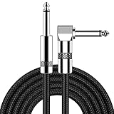
- [Low Noise High Fidelity] New bee guitar cord with 22AWG oxygen-free-copper center conductors provides enhanced signal transfer, the PVC insulation material and the high-density stranded copper double shield provide noise rejection for quiet operation
- [Ideal for Choice] New bee 1/4 straight and right angle metal connectors, ensures a secure connection in any instrument, pedal, or amp jack. L shape great fro on stage guitar stands
- [Wide Compatible] Suitable for mixer, power amplifier, microphone, effector, decoder, equalizer, electoronic organ, electric guitar, electric bass and other electronic equipments. Widely used in the installation of smart home, karaoke speaker, professional stage audio, etc
- [Easy Disassemble] Making incidental repairs quick and easy! Very nice, flexible cable
- [Dont Hesite to Order] 24 hours customer services and professional technology team are standing by
5. Decide whether degradation requires restoration
Identify the limiting failure mode
Restoration is a response to a real acquisition problem: noise that hides cells, blur that merges boundaries, undersampling that removes fine structure, or another demonstrated cause of missed detections. It is not a routine first step for every dataset. More illumination or longer exposure may improve signal, but can increase photodamage, bleaching, or acquisition time; restoration is sometimes considered because those acquisition changes are undesirable.
What Cellpose3 tested
Stringer and Pachitariu’s Cellpose3 paper in Nature Methods, published February 12, 2025, describes denoising, deblurring, and upsampling models trained to improve downstream cellular segmentation rather than only reconstruct pixels. That distinction matters: the objective is useful masks, not necessarily a visually faithful reconstruction.
The paper reports that denoising methods optimized for pixel reconstruction sometimes blurred cell boundaries and reduced segmentation quality. A learned restoration model can also introduce features suggested by its training priors rather than features present in the acquisition. The authors state, “We believe the more-conservative approach should be used whenever possible.” Treat that as a scientific-use principle, not as proof that restoration will help every modality, stain, or cell type.
Quick wins for a faster PC:
Clear out junk files and repair common Windows errorsFree Scan →Scan for outdated or missing drivers - takes under a minuteDriver Scan →Compare raw and restored inputs
Run a controlled comparison on representative images:
| Input state | Useful when | Primary risk | Evidence to collect |
|---|---|---|---|
| Raw acquisition | Boundaries and intensities are already adequate | Noise or blur may cause missed or merged cells | Expert-reviewed masks and failure examples |
| Restored image | Degradation is the demonstrated cause of segmentation errors | Boundaries may shift or invented features may appear | Matched raw/restored review and controls for downstream measurements |
Where feasible, detect cells and generate masks from the restored image, then measure fluorescence, morphology, or other biology on the corresponding raw pixels using those masks. If measurement must be performed on restored pixels, verify the result with suitable matched controls and document the restoration model and settings. Never report a restoration result as a universal accuracy guarantee; Cellpose3’s methods and datasets cover specific tested conditions.
6. Configure channels, diameter, and thresholds empirically
Set the inputs first
Provide the channel and Z-axis settings that match the array you inspected. Confirm that the nuclear or cytoplasmic signal selected in the model interface is the signal actually present at that index. Keep a machine-readable record of these settings with the output masks.
Rank #4

- Digital Family Calendar: BSIMB digital calendar wall touch screen puts an end to your chaotic life. Designed for busy families, the digital calendar keeps you organised and in step with your family. Assign different colours and tasks to each family member to make family scheduling easier and more intuitive.
- Easy Setup and Auto-Sync: Plug in, connect to WLAN and auto-syn your electric calendar in minutes. Digital wall calendar compatible with Google calendar, iCloud calendar, Outlook calendar, Cozi calendar and Yahoo calendar. Add events directly on the device or through the ecalendar app. Electronic calendar stays current whether you're at work or traveling—no manual updates needed.
- All-In-One Home Assistant: Smart calendar builds habits and encourage independence with personalised chores. Display daily meal plan to reduce the daily ‘What's for dinner?’ stress. To-do lists keep track of everything you want. Turning chores into valuable achievements with stars and rewards, making life easier and more enjoyable.
- Manage Anytime Anywhere: BSIMB electronic calendar for family stay connected with the powerful ecalendar app. Access and manage your digital wall calendar anytime, anywhere with free features like stars & rewards, photo screensaver, chore charts, meals plans, to-do lists etc. Electronic planner paid features like Magic Import tailor advanced tools for users with special needs.
- Stay in Touch with Loved Ones: Share photos or videos from your phone to electronic wall calendar with app, instantly update your life moment, and keep in touch with your family, especially those who are far away; you can also surprise your smart calendar digital wall by sending him a wish card; it's also great for keeping tech-illiterate elders in touch with the youngsters.
Tune the parameters people commonly ask about
Cellpose’s FAQ phrases the practical questions as, “What should I set the --flow_threshold/--cellprob_threshold/--diameter parameter to?” Its answer is empirical: “These parameters should be set experimentally by running Cellpose, viewing the results, and tuning the parameters to get the best results.” Start with the model’s suggested values, inspect overlays, and change one setting at a time on a validation subset.
- Diameter: adjust when the expected cell scale differs from the model’s estimate. Check small, typical, and large cells rather than judging one field.
- Cell-probability threshold: raise it when background is being called as cells; lower it only when faint, expert-confirmed cells are being missed.
- Flow threshold: change it when masks have implausible flow or boundary behavior, then inspect whether the change removes real cells as well as artifacts.
These are tuning directions, not universal numerical prescriptions. Keep the image subset, parameter values, software version, and resulting mask set for every comparison.
7. Validate against representative biology
Choose images that expose failure
Validation images should represent the intended experiment, not only the clearest fields. Include variation in cell density, focus, illumination, stain intensity, sample condition, position in the field, and any degradation expected in production. Reserve some images for evaluation rather than repeatedly adjusting settings on every available image.
Use independent expert review
Inspect overlays of masks on the raw acquisition and on any restored input. Look for missed cells, merged cells, splits, boundary shifts, debris called as cells, and systematic errors in particular cell states. Compare model masks with expert annotations made independently of the tuning process. Cellpose’s FAQ advises aiming to meet or exceed human accuracy and suggests estimating human consistency by labeling the same image twice. That repeated labeling is a practical reference for how ambiguous the task itself is; it is not a universal pass threshold.
Separate detection from measurement validation
A model can locate cells acceptably while still altering the boundaries needed for morphology or changing which pixels are included in an intensity measurement. Validate the endpoint you will publish: detection, mask geometry, count, intensity, morphology, or a time-dependent measurement. If segmentation uses restored data and quantification uses raw data, validate that the transferred masks remain appropriate on the raw image.
8. Correct systematic errors and fine-tune carefully
Use a human-in-the-loop cycle
- Run the selected model and settings on a representative batch.
- Accept masks that are correct and edit the errors rather than redrawing every mask unnecessarily.
- Group corrections by failure mode, such as merged touching cells, faint nuclei, or debris.
- Add corrected images and masks to a training set when the same error recurs.
- Evaluate the updated model on images held out from training and from the same intended experiment.
This cycle is useful when errors are systematic, not merely occasional ambiguous cases. A fine-tuned model can memorize fields if the training and evaluation images are too similar, so keep evaluation images independent and representative.
Know when a generalist model is outside its comfort zone
A generalist model is not evidence of performance on every image type. Modality, stain, specimen, cell size, and preparation can differ substantially from the data used to develop a model. Inspect predictions rather than inferring quality from the model name, and plan for expert correction or fine-tuning when the same mistakes recur.
9. Organize files and metadata for reuse
Make filenames interpretable
Use stable identifiers that preserve sample, experiment, position, time point, channel set, and processing state. Keep raw, restored, projected, annotated, and model-output files in distinct locations or with explicit state labels. A DeMemSeg protocol illustrates the value of consistent naming and placing time-point context in filenames; use it as an example of traceability, not a required naming standard.
Record the complete processing context
For each inference run, save:
- source-file identifier and checksum or equivalent file identity;
- array shape, axis order, channel names, and Z or time interpretation;
- data type, bit depth, and any conversion or clipping;
- normalization and restoration operations, including software versions and settings;
- model name and version, diameter, thresholds, channel mapping, and Z handling;
- annotation provenance, reviewer identity or role, and correction history;
- the biological endpoint and whether measurements used raw or restored pixels.
The EMBL-EBI BioImage Archive’s MIFA guidance—Metadata, Incentives, Formats, and Accessibility—was developed to improve reuse of bioimages and annotations for AI applications and is implemented for annotation-file submissions. Consult the current MIFA model reference for exact fields; the practical goal is that another researcher can understand what each pixel and mask represents without opening the original microscope software.
The Tool Desk
Outbyte Driver Updater FREEFix the driver behind crashes, sound loss and screen glitchesFind Drivers →Outbyte PC Repair FREEClear out junk files and repair common Windows errorsFree Scan →10. A reproducible preparation checklist
- Original acquisition is retained unchanged.
- Source and working-copy identifiers are linked.
- Y, X, channel, Z, and time axes are explicitly documented.
- Cellpose input layout is one of the supported arrangements and matches the configured axes.
- Nuclear, cytoplasmic, membrane, and other channels are mapped by index and name.
- Bit depth, data type, clipping, and percentile range were checked after conversion.
- Built-in model normalization was reviewed before any external normalization.
- Every restoration or projection step has a stated reason and saved setting.
- Raw and transformed inputs were compared on representative images.
- Segmentation and biological quantification use the intended input states.
- Diameter and threshold parameters were tuned by inspecting results, not copied as universal values.
- Independent expert review covers the variation expected in the experiment.
- Corrections, fine-tuning data, held-out evaluation images, and software versions are retained.
- Metadata and annotation files follow an interpretable, reusable organization.
Common problems and what to check first
| Symptom | Likely preparation issue | First corrective action |
|---|---|---|
| All cells appear in the wrong size or orientation | Incorrect axis order or diameter assumption | Reinspect the array shape, set channel and Z axes explicitly, and retune diameter on representative fields. |
| One channel appears empty or the wrong stain drives the masks | Channel mapping changed during export | Compare channel indices and names in the source and working files; recreate the mapping explicitly. |
| Background is segmented as cells | Thresholds, normalization, or clipping changed the intensity distribution | Review the model’s built-in normalization, inspect clipped pixels, and tune the cell-probability threshold experimentally. |
| Touching cells merge after denoising | Restoration blurred boundaries | Compare with the raw input and test a less aggressive or different restoration; validate masks against expert labels. |
| Restored images look sharper but measurements change | Learned restoration altered boundaries or intensities | Use restored data for detection only where justified and measure on raw pixels, or establish matched controls for restored measurements. |
| Results work on one field but fail on another sample | Distribution mismatch or overfitting during tuning | Evaluate on held-out, representative images and add expert corrections or fine-tune with independent validation. |
When local hardware is not enough
Cellpose’s FAQ describes a remote workflow for users without suitable local GPU capability: transfer images to a remote machine, run a pretrained model, annotate masks, and use a hosted notebook such as Colab or a cluster. The computing location does not remove the preparation obligations. Transfer only data permitted by your governance rules, preserve the same channel mapping and transforms, and download masks together with the run settings and source identifiers.
The practical standard for a trustworthy result
A trustworthy AI cell-analysis input is not the most heavily processed image. It is an image whose axes, channels, values, transformations, model settings, and biological purpose are explicit and reproducible. Use restoration only when a measured degradation justifies it, inspect the resulting masks against independent human work, and keep quantitative biology tied to the raw acquisition whenever possible. This approach lets you improve detection without losing track of what the microscope actually measured.
Quick Recap
Product prices and availability are accurate as of the date/time indicated and are subject to change. Any price and availability information displayed on Amazon at the time of purchase will apply.

