A microscopy manuscript should let a reader trace every published panel back to an unaltered acquisition file. Preserve the original files, work on copies, record the microscope and software context, list processing operations in order, identify adjustments that affect interpretation, and explain composites in the Methods and figure legends. Quantitative measurements must remain tied to appropriate raw data rather than a visually enhanced display image.
Start with a traceable image record
Image-processing documentation is not a description of how a figure looks. It is a chain of custody for the data: which specimen and acquisition produced the source file, which software opened it, which operations were applied, which version became a figure panel, and which file was used for measurement. The community checklist for quantitative light-microscopy publication treats this chain as part of complete reporting, not as optional production detail.

As an Amazon Associate I earn from qualifying purchases.
Keep the original, unaltered acquisition files in a protected location. Do not overwrite them when adjusting brightness, registering channels, making projections, or exporting a figure. Create working copies and preserve a clear relationship between each source file, each intermediate file, and each final panel. Institutional guidance from KU Leuven and the Office of Research Integrity (ORI) both make retention of the original and processing on a copy central requirements.
Quick wins for a faster PC:
Fix the driver behind crashes, sound loss and screen glitchesFind Drivers →Clear out junk files and repair common Windows errorsFree Scan →Scan for outdated or missing drivers - takes under a minuteDriver Scan →Use stable identifiers
Give every acquisition a stable identifier that survives renaming and figure assembly. A useful identifier can combine the experiment, specimen, field, and acquisition date, such as EXP07_S03_F12_2026-04-18. Keep the microscope-generated filename in the record as well; do not rely on a new descriptive filename alone.
#1 Best Overall

- [Ultra-Lightweight Ear Buds Designed for Small Ears] Each earbud weighs only 3.7g and features a compact, ergonomic in-ear design made especially for small ears. Secure, low-profile, and comfortable for workouts, all-day wear or overnight sleeping.The ultra-slim profile sits flush in your ear with no hard edges poking or rubbing, so you can even sleep on your side without discomfort.
- [Immersive Stereo Sound with TOZO OrigX Technology] TOZO OrigX tuning delivers clear vocals, balanced mids, and natural stereo sound for music, podcasts, and videos.
- [Long Battery Life for Daily Use] Get up to 7 hours of playtime on a single charge, with up to 32 hours total using the charging case—ideal for workdays, commuting, and extended listening sessions.
- [Bluetooth 5.3 & Stable Connection] Bluetooth 5.3 provides fast pairing, stable wireless performance, and reduced dropouts as you move around home or office.
- [Deep Bass with Clear Vocals] High-performance drivers produce punchy bass while keeping vocals clean and detailed for everyday listening.
For each output, record its immediate parent file. A simple manifest can contain these columns:
- Source-file identifier and original filename
- Specimen, preparation, condition, and field or time point
- Acquisition date and operator
- Processing software and version
- Operation, parameters, and order
- Output filename and destination figure or supplementary item
- Whether the output was used for display, measurement, or both
If a laboratory uses an electronic notebook, link the manifest entry to the notebook page or analysis project. If it uses plain files, keep a read-only copy of the manifest with the original data and another copy with the submission archive.
Record the acquisition context before describing processing
Processing cannot be interpreted without knowing how the image was acquired. Include the acquisition details that determine resolution, signal, intensity, color, and scale. The American Society for Biochemistry and Molecular Biology (ASBMB) checklist identifies the following information as relevant for light-microscopy publication.
| Context | What to record | Why it matters |
|---|---|---|
| Microscope | Make and model; imaging modality where relevant | Different optical systems and detectors produce different sampling and contrast characteristics. |
| Objective | Type, magnification, and numerical aperture | These values affect resolution, collection efficiency, and the scale represented by each pixel. |
| Environment | Temperature and imaging medium | Environmental conditions can alter specimen behavior, refractive-index matching, and signal stability. |
| Labels | Fluorochromes or stains, including the channel associated with each label | Readers need to know what each displayed channel represents. |
| Detector | Camera make and model, or the detector type used | Detector characteristics influence sensitivity, dynamic range, and noise. |
| Capture software | Acquisition software name and version when available | The software can determine file metadata, bit depth, deconvolution options, and export behavior. |
| File properties | Original format, dimensions, bit depth, channels, z-spacing, and time interval where applicable | These properties establish what information was present before processing. |
| Scale | Pixel size or voxel size and a scale bar on the figure | A reader must be able to relate displayed distances to physical dimensions. |
Wiley’s guidance also calls for sample preparation, stains, microscope, camera, and capture or editing software in the Methods or figure legend. Put general acquisition information in Methods, then repeat only figure-specific differences—such as a different objective, exposure, or channel—in the relevant legend.
Include acquisition settings that affect interpretation
When the setting is consequential, record it rather than hiding it behind the instrument name. Examples include exposure or dwell time, excitation and emission configuration, laser power, detector gain, binning, pixel size, z-step, number of averaged frames, and whether the detector reached saturation. The cited checklists specifically require the core hardware and software context; adding the settings that control signal and sampling makes the record more reproducible and helps explain why two images should or should not be compared.
State whether images intended for comparison were acquired under identical conditions. ORI advises that compared images be acquired identically and that intensity measurements use raw data calibrated to a known standard. If conditions differed, identify the difference and explain how the analysis accounts for it; do not present differently acquired images as though their intensities were directly interchangeable.
Rank #2

- 【IP68 Waterproof Multifunctinal Smart Watch】Torjalph fitness activity tracker smart watch is IP68 waterproof technology ,you can wear it when swimming,handwashing,it's rain-proof which means that you perform multiple exercise modes outside without worrying about sweat or rain corroding your fitness smart watch,and this watch equipped with a 230 mAh large capacity battery magnetic charging ,and this sports activity tracker watch can be used 5-7 days If fully charged.
- 【Larger 1.83'' Full Screen Watch & Multi-dial Selection】1.83 inch full HD color touch screen smart watch for men / women provides excellent 240*284 resolution image quality ,This Bluetooth smart watch offers a better touch experience and remarkable immersion and ,more than 200 watch faces for you to choose from App"Da Fit ", you can change the dial style according to your mood to wear keeping the world on your wrist
- 【Accurate Activity Tracker Smart Watch 】sports watch activity tracker uses sweat and dust resistant breathable silicone strap, our Fitness Tracker Smart Watch for Android iOS Phones supports 12 sports modes,walking,running,bicycle,swimming,badminton,basketball,football,rope skipping. This bluetooth calls smart watch for men women is lightweight and stylish, bringing more convenient and relaxing sports experience while also providing more protection.
- 【Your 24 Hour Personal Daily Health Monitor】Fitness watch for women men full touch screen IP67 waterproof bluetooth smartwatch for Android iOS Phones with heart rate blood pressure monitor multifunction Andriod smart watch with steps counter sleep monitor sports fitness watch, monitoring blood oxygen saturation allows you to understand your physical condition in real time to track fitness progress ,to know your health status in time.
- 【More Functions Bluetooth Call Smart Watch】This fitness activity smart watch full screen for iPhone and Android phones includes a bunch of new features including:The bluetooth smart watch can hang up the phone, receive and display WhatsAPP /Text /Facebook /Twitter /Messenger reminder.Support sedentary reminder, alarm clock, stopwatch, timer,weather information,music control, remote camera control compatible with Android 5.0 and above or iOS 9.0 and above smartphones.
Describe the processing sequence, not just the final appearance
Write the workflow in chronological order. A statement such as “images were processed” is too vague because it omits the operations that can change interpretation. Name the software used for acquisition and every post-acquisition package that made a consequential change. Then state what each operation did, its important parameters, and its purpose.
PC Slower Than It Used to Be?
A free scan shows the junk files, broken settings and background clutter dragging Windows down - then fixes them in one click.Free scan · Windows 10 & 11Outdated Drivers Are Slowing You Down
One free scan finds every outdated or missing driver and matches the right update for your exact hardware.Free scan · exact hardware matchSeparate display operations from analytical operations
Readers need to know whether an operation changed only how pixels are shown or changed the data used for measurement. Organize the record into two paths when appropriate:
- Display path: operations used to make a figure legible, such as a whole-image linear contrast adjustment, channel coloring, or a projection made solely for presentation.
- Analysis path: operations used to calculate intensity, area, volume, object count, colocalization, or another measurement.
If one output served both purposes, say so and explain which data values entered the calculation. A display export in a presentation format should not silently become the measurement source.
List operations and parameters in order
Use a numbered sequence that another researcher could reproduce. Depending on the experiment, the sequence might include:
- Import the original file without changing its bit depth or channel order.
- Correct a documented acquisition artifact, such as uneven illumination, and identify the correction image or algorithm.
- Register channels or time points, specifying the reference channel and transformation method.
- Apply deconvolution, naming the software, algorithm, point-spread-function source, iteration or stopping rule, and whether the result was used for display, analysis, or both.
- Generate a maximum-intensity, average-intensity, or other projection, stating the z-range and projection type.
- Segment objects with a stated thresholding or classifier procedure, including whether thresholds were fixed, manually chosen, or calculated per image.
- Measure objects or regions from the defined data set and export the values with the analysis settings.
- Render a three-dimensional surface or volume, documenting the smoothing, threshold, opacity, and lighting choices that could affect visual interpretation.
- Export a figure at the journal’s required format and resolution without resampling away information needed for interpretation.
Not every manuscript needs every parameter, but every operation that could affect a reader’s interpretation or a reported number does. Record the actual sequence, even when the software presents operations in a different order during interactive work.
Name consequential transformations
Explicitly identify operations such as deconvolution, 3D reconstruction, surface or volume rendering, gamma adjustment, thresholding, background subtraction, denoising, sharpening, channel registration, stitching, projection, segmentation, and object exclusion. Explain why the operation was used and whether it was applied to the entire image, to all samples, or only to a defined analysis region.
Rank #3
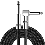
- [Low Noise High Fidelity] New bee guitar cord with 22AWG oxygen-free-copper center conductors provides enhanced signal transfer, the PVC insulation material and the high-density stranded copper double shield provide noise rejection for quiet operation
- [Ideal for Choice] New bee 1/4 straight and right angle metal connectors, ensures a secure connection in any instrument, pedal, or amp jack. L shape great fro on stage guitar stands
- [Wide Compatible] Suitable for mixer, power amplifier, microphone, effector, decoder, equalizer, electoronic organ, electric guitar, electric bass and other electronic equipments. Widely used in the installation of smart home, karaoke speaker, professional stage audio, etc
- [Easy Disassemble] Making incidental repairs quick and easy! Very nice, flexible cable
- [Dont Hesite to Order] 24 hours customer services and professional technology team are standing by
For nonlinear transformations, give special care to the disclosure. Gamma changes and other nonlinear mappings alter the relationship between the displayed value and the underlying intensity. KU Leuven states that nonlinear adjustments must be disclosed in the figure legend or represented clearly in a quantitative color bar. A legend should name the transformation rather than describing it as a generic “contrast enhancement.”
Apply display adjustments without hiding information
Whole-image brightness, contrast, or color adjustments can be acceptable when they are applied consistently and do not conceal or misrepresent information. “Whole-image” means the adjustment is not selectively applied to one object, cell, band, or region to create a desired result. State whether the same display range or transformation was used for all images being compared. If ranges differ for visibility, disclose that difference and avoid implying that color or brightness is quantitatively comparable.
Adjustments that require explicit disclosure
- Nonlinear mappings such as gamma, logarithmic, or sigmoid transformations
- Clipping of low or high values
- Background subtraction or flat-field correction
- Deconvolution, denoising, sharpening, or smoothing
- Color changes that map channels to a different visual representation
- Projection, resampling, interpolation, or rotation that changes spatial representation
- Thresholding or segmentation used to define what is visible or measurable
Keep a copy of the image before and after each consequential step, or retain a reproducible processing script or recorded command history. A screenshot of a slider is not a sufficient record if the software can save exact parameters or a macro.
Recommended Free Tools
Do not alter individual features
The KU Leuven guidance is direct: “No specific feature within an image may be enhanced, obscured, moved, removed, or introduced.” Do not erase debris, copy a signal into a blank region, move an object, clone a cell, or selectively sharpen a structure. If an artifact must be removed for a clearly labeled illustration, do not use that altered image as evidence for a result, and identify the illustration as such.
Make composites and multi-panel figures unambiguous
A composite can combine channels, fields, z-planes, time points, or separate exposures. It is acceptable only when the construction is made clear. State which source images were combined, whether they were acquired from the same field and under the same conditions, and which operations were applied to each component.
Mark boundaries and source relationships
If separate fields or exposures are placed side by side, show the boundary or spacing clearly and label each component. If a crop is used, indicate that it is a crop and identify the parent image. For a merged-channel panel, provide the individual channels when they are necessary to interpret overlap. A legend should distinguish a true simultaneous acquisition from a montage assembled from separate acquisitions.
Rank #4

- Digital Family Calendar: BSIMB digital calendar wall touch screen puts an end to your chaotic life. Designed for busy families, the digital calendar keeps you organised and in step with your family. Assign different colours and tasks to each family member to make family scheduling easier and more intuitive.
- Easy Setup and Auto-Sync: Plug in, connect to WLAN and auto-syn your electric calendar in minutes. Digital wall calendar compatible with Google calendar, iCloud calendar, Outlook calendar, Cozi calendar and Yahoo calendar. Add events directly on the device or through the ecalendar app. Electronic calendar stays current whether you're at work or traveling—no manual updates needed.
- All-In-One Home Assistant: Smart calendar builds habits and encourage independence with personalised chores. Display daily meal plan to reduce the daily ‘What's for dinner?’ stress. To-do lists keep track of everything you want. Turning chores into valuable achievements with stars and rewards, making life easier and more enjoyable.
- Manage Anytime Anywhere: BSIMB electronic calendar for family stay connected with the powerful ecalendar app. Access and manage your digital wall calendar anytime, anywhere with free features like stars & rewards, photo screensaver, chore charts, meals plans, to-do lists etc. Electronic planner paid features like Magic Import tailor advanced tools for users with special needs.
- Stay in Touch with Loved Ones: Share photos or videos from your phone to electronic wall calendar with app, instantly update your life moment, and keep in touch with your family, especially those who are far away; you can also surprise your smart calendar digital wall by sending him a wish card; it's also great for keeping tech-illiterate elders in touch with the youngsters.
Do not imply continuity where none exists. A stitched panorama, tiled acquisition, or montage should be labeled as such; a blank separator or thin line can help show the join. For a time series, label the time points and state the interval. For a z-stack projection, state the z-range and projection method rather than presenting the projection as a single optical section.
Keep scale bars tied to the correct panel
Use a scale bar derived from the pixel or voxel dimensions of the source image. If panels have different magnifications, crops, or resampling factors, give each panel its own scale bar or state exactly which panels share one. Do not resize a figure panel after adding a scale bar unless the bar is regenerated or scaled with the image.
Independent reader supportYour contribution helps us test, update, and keep practical guides available for everyone.Keep quantitative analysis faithful to the data
Visual plausibility is not a substitute for quantitative integrity. ORI advises that intensity measurements be performed on raw data and calibrated to a known standard. In practice, preserve the original numerical values and document any calibration, background correction, normalization, or unit conversion applied before measurement.
Define which files enter the analysis
State whether measurements came from original files, lossless converted files, or processed copies. If a correction was necessary, identify the correction and explain why it does not invalidate the measurement. Do not measure a screenshot, a compressed image, or a rendered surface when the reported value is supposed to represent raw intensity or area.
State selection and exclusion criteria
Explain how fields, cells, particles, or time points were selected. Criteria should be defined before or independently of the desired result where possible. Report exclusions, failed acquisitions, saturated images, and missing data rather than silently removing inconvenient examples. The ASBMB checklist calls for image-selection and analysis criteria, replicates, and the statistical approach relevant to the analysis.
The Tool Desk
Outbyte PC Repair FREERepair Windows errors before they cause bigger problemsFix Now →Outbyte Driver Updater FREEFix the driver behind crashes, sound loss and screen glitchesFind Drivers →Match processing across groups
Apply the same analysis pipeline and parameter rules to compared groups unless a justified, documented difference is part of the design. If a threshold is selected separately for each image, state that rule and explain how it affects comparability. Keep the analysis settings, masks, and output tables with the source identifiers so a reader can distinguish biological variation from processing variation.
Place details where readers will find them
Use the Methods for the general workflow and the figure legend for operations specific to a displayed figure. A concise Methods paragraph can identify the software and the standard sequence; the legend can then state the channel merge, crop, projection, display range, nonlinear mapping, or composite construction used for that panel.
Methods example
“Images were acquired on a [microscope make and model] using a [objective type, magnification, and numerical aperture] objective and a [camera or detector]. Samples were maintained at [temperature] in [imaging medium] and labeled with [fluorochromes or stains]. Acquisition used [software and version], with images stored in the original format and bit depth. Working copies were processed in [post-acquisition software and version]. The sequence was [operation 1], [operation 2], and [operation 3], using the parameters described in Supplementary Table [identifier]. Quantitative intensity measurements were made from the unaltered acquisition data after [calibration or documented correction].”
Replace each bracketed item with the actual value before submission; do not leave template markers in a manuscript. If a detail does not apply, omit it rather than inserting an inapplicable value.
What’s actually slowing this PC down?
Pick the symptom - the matching free tool is one click away.
Figure-legend example
“Representative maximum-intensity projection of the z-range [range]. All channels were displayed with the same linear intensity range within the comparison. A gamma adjustment of [value or software setting] was applied to the displayed image only. The merged panel combines channels acquired from the same field; individual channels are shown at left. The white scale bar is [length].”
For a composite of separate fields, replace the same-field statement with a precise description of the assembly and mark the boundaries in the figure. For a rendered surface or volume, name the rendering method and the threshold or opacity rule that materially affects what is visible.
Follow the destination journal’s current rules
There is no single universal image-submission rule. The target journal controls file formats, resolution, color mode, source-data requirements, figure-legend wording, and retention expectations. Check those instructions immediately before submission and again if the manuscript is transferred to another journal.
Put workflow details in Methods and figure-specific operations in legends unless the journal specifies another location. Retain the original files, processing records, analysis outputs, and the submitted figure package in a form that can be retrieved if editors request clarification. Wiley advises retaining original unadjusted data for five years after publication; that is Wiley’s recommendation, not a universal retention period for every journal.
Do these 3 things before closing this tab:
1Repair Windows errors before they cause bigger problems2Scan for outdated or missing drivers - takes under a minute3Clear out junk files and repair common Windows errorsCheck technical requirements separately from disclosure
A journal may require a particular file format or resolution for publication while also asking for source data in a different format. Converting a file for production does not replace preservation of the original. Keep both the source and the submission derivative, and record the conversion so the relationship is clear.
Submission checklist
- Original unaltered acquisition files are preserved and protected from overwrite.
- Every published panel and analysis output can be traced to a source identifier.
- Microscope, objective, numerical aperture, temperature, medium, labels, camera or detector, and acquisition software are recorded.
- File format, bit depth, pixel or voxel dimensions, and scale information are available.
- Post-acquisition software and versions are named.
- Processing operations appear in chronological order with consequential parameters.
- Display-only adjustments are distinguished from analysis operations.
- Nonlinear mappings, clipping, projection, rendering, thresholding, and other interpretive transformations are disclosed.
- No individual feature has been selectively enhanced, obscured, moved, removed, or introduced.
- Compared images were acquired and processed under consistent conditions, or differences are explained.
- Intensity measurements use appropriate raw data and documented calibration.
- Selection criteria, exclusions, replicates, and statistical methods are stated.
- Composite boundaries, crops, channel identities, time points, and z-ranges are clear.
- Scale bars match the final displayed dimensions.
- The target journal’s current file, source-data, resolution, and retention rules have been checked.
Common failures and how to repair them
| Failure | Why it is a problem | Repair before submission |
|---|---|---|
| “Images were processed” with no detail | The reader cannot tell what changed or reproduce the workflow. | Name each software package and list operations in order with relevant parameters. |
| Only the final JPEG or raster figure remains | Compression and rendering may have discarded numerical information. | Recover and preserve the original acquisition files; identify which outputs were made from them. |
| A single cell or band was selectively brightened | The alteration can misrepresent the underlying observation. | Restore the unaltered panel and apply any display adjustment to the complete image, or omit the adjustment. |
| Gamma or clipping is not mentioned | Displayed brightness no longer has an obvious relationship to intensity. | Disclose the nonlinear mapping or clipping in the legend and keep quantitative analysis on suitable raw data. |
| A montage looks like one continuous field | Readers may infer spatial continuity or a common exposure that does not exist. | Mark boundaries and explain the source fields, exposures, and assembly. |
| Measurements were made from a rendered or screenshot image | Rendering and export can change values and spatial relationships. | Re-run measurements from the original or a documented, lossless analysis copy. |
| Different groups use different thresholds without explanation | Processing differences can become a hidden source of group differences. | Define a consistent rule or justify and report the group-specific procedure. |
| Scale bars were added before resizing | The bar may no longer represent the displayed distance. | Regenerate the bar after final resizing or scale it with the panel. |
A well-documented microscopy figure does not require every intermediate image to appear in the paper. It does require that the originals, processing history, analytical source, and display changes remain identifiable, and that the figure itself gives readers enough information to interpret what they see.
Quick Recap
Product prices and availability are accurate as of the date/time indicated and are subject to change. Any price and availability information displayed on Amazon at the time of purchase will apply.

