Quick wins for a faster PC:
Clear out junk files and repair common Windows errorsFree Scan →Fix the driver behind crashes, sound loss and screen glitchesFind Drivers →Repair Windows errors before they cause bigger problemsFix Now →Short answer: you cannot confirm an AI highlight by looking at the colored outline alone. It is a model-generated cue, not proof of an abnormality. The safe check is to view the marked location on the corresponding original scan images, then ask the clinician interpreting the study to explain whether it matches the written report and is confirmed on the source images.
A screenshot may show where software placed a prediction but hide the image series, measurements, viewing controls, and coded result details needed for meaningful review. Treat the highlight as a prompt for a clinical conversation, not as a diagnosis you can verify at home.

As an Amazon Associate I earn from qualifying purchases.
What an AI highlight actually represents
Medical-imaging software analyzes pixels from a particular study, series, and acquisition protocol. Its output may be a contour, arrow, heat map, bounding box, measurement, or other annotation. That graphic shows where the algorithm placed a prediction or attention. It does not independently establish that a lesion, fracture, clot, nodule, or other abnormality exists.
The Tool Desk
Outbyte PC Repair FREEClear out junk files and repair common Windows errorsFree Scan →Outbyte Driver Updater FREEScan for outdated or missing drivers - takes under a minuteDriver Scan →Whether the prediction is useful depends on the exact task, software version, patient and study matching, image quality, and the clinician’s interpretation. A color, shape, opacity, or confidence score has no universal meaning. Only the documentation for that specific system can define those conventions.
#1 Best Overall

- [Ultra-Lightweight Ear Buds Designed for Small Ears] Each earbud weighs only 3.7g and features a compact, ergonomic in-ear design made especially for small ears. Secure, low-profile, and comfortable for workouts, all-day wear or overnight sleeping.The ultra-slim profile sits flush in your ear with no hard edges poking or rubbing, so you can even sleep on your side without discomfort.
- [Immersive Stereo Sound with TOZO OrigX Technology] TOZO OrigX tuning delivers clear vocals, balanced mids, and natural stereo sound for music, podcasts, and videos.
- [Long Battery Life for Daily Use] Get up to 7 hours of playtime on a single charge, with up to 32 hours total using the charging case—ideal for workdays, commuting, and extended listening sessions.
- [Bluetooth 5.3 & Stable Connection] Bluetooth 5.3 provides fast pairing, stable wireless performance, and reduced dropouts as you move around home or office.
- [Deep Bass with Clear Vocals] High-performance drivers produce punchy bass while keeping vocals clean and detailed for everyday listening.
Overlays can make a model’s output easier to inspect, but a strong statistical result does not automatically translate into reliable care. A Radiology editorial explains that a high area-under-the-curve value can coexist with an operating point that misses many abnormalities; the example is an illustration of why clinical context matters, not an accuracy rate for every model. Read the Radiology editorial’s discussion of evaluating imaging-AI studies.
First determine whether you have a screenshot or an inspectable result
The format determines what you can examine, but it never guarantees that the highlighted area is correct.
| What you have | What it usually contains | What you can reasonably inspect | Main limitation |
|---|---|---|---|
| Static annotated image (DICOM secondary capture) | A frozen image with an outline, label, or other annotation burned into the displayed pixels | The approximate marked location and the visible anatomy in that single view | You may be unable to scroll through the source series, change window or level, or access machine-readable measurements and findings |
| Structured, interactive AI result | A link to the source DICOM images plus encoded locations, measurements, segmentations, or qualitative findings | The same location across slices, the original image settings, and any supplied measurements or labels | More information is available, but the result still needs clinical interpretation and can still be wrong |
The RSNA primer on the IHE AI Results (AIR) profile describes how pixel-based results can be stored, retrieved, and displayed with the corresponding DICOM images. It distinguishes a static secondary capture from structured reports and segmentations that preserve locations, measurements, and coded findings. See the RSNA primer on AI results and radiology workflow.
What’s actually slowing this PC down?
Pick the symptom - the matching free tool is one click away.
Clues that an image is only a static capture
- There is one flattened image rather than a stack you can scroll through.
- The outline is part of the pixels and cannot be hidden or selected.
- No series, slice, measurement, or finding fields are available.
- Window/level, zoom, pan, and other viewer controls do not work.
Clues that a result is interactive
- The annotation can be toggled, selected, or hidden.
- The viewer can jump to the marked slice or display the source series beside the result.
- A measurement, location, or finding is presented as a separate data element.
- The system identifies the study, series, and image frame associated with the result.
These clues describe inspectability, not correctness. A beautifully integrated viewer can display a false positive, while a static image can happen to show a real finding.
Rank #2

- 【IP68 Waterproof Multifunctinal Smart Watch】Torjalph fitness activity tracker smart watch is IP68 waterproof technology ,you can wear it when swimming,handwashing,it's rain-proof which means that you perform multiple exercise modes outside without worrying about sweat or rain corroding your fitness smart watch,and this watch equipped with a 230 mAh large capacity battery magnetic charging ,and this sports activity tracker watch can be used 5-7 days If fully charged.
- 【Larger 1.83'' Full Screen Watch & Multi-dial Selection】1.83 inch full HD color touch screen smart watch for men / women provides excellent 240*284 resolution image quality ,This Bluetooth smart watch offers a better touch experience and remarkable immersion and ,more than 200 watch faces for you to choose from App"Da Fit ", you can change the dial style according to your mood to wear keeping the world on your wrist
- 【Accurate Activity Tracker Smart Watch 】sports watch activity tracker uses sweat and dust resistant breathable silicone strap, our Fitness Tracker Smart Watch for Android iOS Phones supports 12 sports modes,walking,running,bicycle,swimming,badminton,basketball,football,rope skipping. This bluetooth calls smart watch for men women is lightweight and stylish, bringing more convenient and relaxing sports experience while also providing more protection.
- 【Your 24 Hour Personal Daily Health Monitor】Fitness watch for women men full touch screen IP67 waterproof bluetooth smartwatch for Android iOS Phones with heart rate blood pressure monitor multifunction Andriod smart watch with steps counter sleep monitor sports fitness watch, monitoring blood oxygen saturation allows you to understand your physical condition in real time to track fitness progress ,to know your health status in time.
- 【More Functions Bluetooth Call Smart Watch】This fitness activity smart watch full screen for iPhone and Android phones includes a bunch of new features including:The bluetooth smart watch can hang up the phone, receive and display WhatsAPP /Text /Facebook /Twitter /Messenger reminder.Support sedentary reminder, alarm clock, stopwatch, timer,weather information,music control, remote camera control compatible with Android 5.0 and above or iOS 9.0 and above smartphones.
A safe, step-by-step way to check the match
- Identify the exact study. Confirm the patient, body part, laterality, modality, and examination date. An AI result must correspond to the same study and series that the clinician is interpreting.
- Ask for the original images, not only the marked screenshot. Request that the clinician show the source images in the diagnostic viewer. Do not rely on a phone image, a portal thumbnail, or a printout as the complete examination.
- Locate the marked slice or region. Ask which image number, series, plane, or anatomical landmark contains the highlighted area. If the result is structured, the viewer may be able to jump directly to that location.
- Review neighboring images. A suspected finding may extend across several slices or appear differently in another plane. The clinician can scroll and adjust the display appropriately; a single frozen frame cannot establish that context.
- Compare the annotation with the anatomy. Ask what structure the outline is touching and whether the shape, density, signal, or enhancement is visible on the original images without the overlay. This is a request for explanation, not an instruction to diagnose yourself.
- Check any supplied measurement. If the software reports a size, volume, or other value, ask how it was measured and whether the clinician accepts it. A machine-readable number is easier to review than a burned-in graphic, but it is not automatically accurate.
- Match the location to the written report. Ask whether the radiology report describes the same side, organ, level, and finding. If the report uses different wording, ask the interpreting clinician to reconcile the terms.
- Ask for a clinical conclusion. The clinician should state whether the highlighted area is confirmed, represents a benign or expected structure, is an artifact, or needs additional evaluation. Do not make that determination from the overlay alone.
The U.S. Food and Drug Administration’s documentation for one cleared image viewer describes displaying annotations and measurements over DICOM and AI/CAD images and emphasizes that professionals must ensure appropriate viewing conditions and image quality for the clinical purpose. That is a product-specific description, not a guarantee about every viewer. Read the FDA documentation for the OmegaAI Image Viewer (K222476).
Questions that make the clinician’s explanation specific
Bring the image and report together and ask concise, location-focused questions:
- “Which image or slice shows the finding?”
- “How does the highlighted location correspond to the wording in the report?”
- “Is the highlighted area confirmed on the original images?”
- “Does the annotation cover the whole finding, or is it only a pointer?”
- “What measurement did the system provide, and do you agree with it?”
- “Could this be normal anatomy, motion, noise, or another artifact?”
- “Was this AI tool used for this exact body part and examination type?”
- “Does the finding change what happens next?”
These questions are designed to connect the software output to the images and report. They do not turn a patient into an independent image reader.
Recommended Free Tools
Why the original image series matters
Radiologists interpret examinations as series rather than isolated pictures. They may compare multiple slices, planes, phases, sequences, prior studies, and display settings. A highlight can appear to sit over a structure because of projection, motion, partial-volume effects, or an incorrect registration. Scrolling through the source images helps reveal whether the marked area persists and where it is anatomically located.
Rank #3
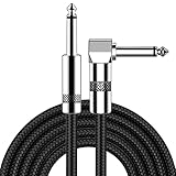
- [Low Noise High Fidelity] New bee guitar cord with 22AWG oxygen-free-copper center conductors provides enhanced signal transfer, the PVC insulation material and the high-density stranded copper double shield provide noise rejection for quiet operation
- [Ideal for Choice] New bee 1/4 straight and right angle metal connectors, ensures a secure connection in any instrument, pedal, or amp jack. L shape great fro on stage guitar stands
- [Wide Compatible] Suitable for mixer, power amplifier, microphone, effector, decoder, equalizer, electoronic organ, electric guitar, electric bass and other electronic equipments. Widely used in the installation of smart home, karaoke speaker, professional stage audio, etc
- [Easy Disassemble] Making incidental repairs quick and easy! Very nice, flexible cable
- [Dont Hesite to Order] 24 hours customer services and professional technology team are standing by
Structured segmentations and reports can preserve the coordinates, measurements, and qualitative findings in a form that a viewer or another system can process. A secondary capture preserves only the appearance of the screen at one moment. If all you received is a flattened image, ask the imaging facility or clinician to interpret the underlying examination rather than trying to reconstruct it from the screenshot.
How to judge claims about an AI system’s performance
No universal percentage tells you how often every AI highlight is correct. Performance is task-specific and can change when the patient population, scanner, acquisition protocol, prevalence of disease, or clinical workflow changes.
| Evidence detail | Why it matters to the highlight you are viewing |
|---|---|
| Reference standard | The comparison target used to label cases, such as expert consensus, pathology, follow-up, or another defined standard. A vague “ground truth” label makes the basis of the result harder to judge. |
| Internal versus external testing | Testing on held-out data from the development setting is different from testing on an entirely external dataset. External performance gives information about generalization to a different population or site. |
| Image-acquisition protocol | Scanner type, sequence, contrast phase, slice thickness, and other protocol details can affect whether a model behaves as expected. |
| Intended clinical task | A tool designed to flag suspected pulmonary emboli, for example, is not a general detector for every chest abnormality. |
| Operating point and errors | Sensitivity, specificity, false positives, false negatives, and the chosen threshold describe different trade-offs. One summary metric can conceal clinically important misses. |
The 2024 CLAIM update recommends reporting the data source, study goal, acquisition protocol, and reference standard. It also distinguishes internal from external testing and discourages using the word “validation” without explaining what was actually tested. Read the CLAIM 2024 reporting checklist. Those reporting details help you understand a published claim; they do not validate an individual patient’s result.
PC Slower Than It Used to Be?
A free scan shows the junk files, broken settings and background clutter dragging Windows down - then fixes them in one click.Free scan · Windows 10 & 11Crashes, No Sound, or Screen Glitches?
Random freezes, missing sound and display glitches usually trace back to one bad driver. Find and replace yours safely.Free scan · under a minuteWhy a confidence score is not a verdict
A score is meaningful only within the model, task, calibration, and threshold that produced it. A higher number may indicate stronger model confidence, but it does not prove that the marked structure is abnormal or that the system is operating correctly on your examination. Ask the interpreting clinician how, if at all, the score is used in that workflow.
Rank #4

- Digital Family Calendar: BSIMB digital calendar wall touch screen puts an end to your chaotic life. Designed for busy families, the digital calendar keeps you organised and in step with your family. Assign different colours and tasks to each family member to make family scheduling easier and more intuitive.
- Easy Setup and Auto-Sync: Plug in, connect to WLAN and auto-syn your electric calendar in minutes. Digital wall calendar compatible with Google calendar, iCloud calendar, Outlook calendar, Cozi calendar and Yahoo calendar. Add events directly on the device or through the ecalendar app. Electronic calendar stays current whether you're at work or traveling—no manual updates needed.
- All-In-One Home Assistant: Smart calendar builds habits and encourage independence with personalised chores. Display daily meal plan to reduce the daily ‘What's for dinner?’ stress. To-do lists keep track of everything you want. Turning chores into valuable achievements with stars and rewards, making life easier and more enjoyable.
- Manage Anytime Anywhere: BSIMB electronic calendar for family stay connected with the powerful ecalendar app. Access and manage your digital wall calendar anytime, anywhere with free features like stars & rewards, photo screensaver, chore charts, meals plans, to-do lists etc. Electronic planner paid features like Magic Import tailor advanced tools for users with special needs.
- Stay in Touch with Loved Ones: Share photos or videos from your phone to electronic wall calendar with app, instantly update your life moment, and keep in touch with your family, especially those who are far away; you can also surprise your smart calendar digital wall by sending him a wish card; it's also great for keeping tech-illiterate elders in touch with the youngsters.
When the highlight and the report do not seem to agree
Do not choose between them yourself. First verify that the overlay belongs to the same patient, study, series, and software run. A mismatch can result from selecting the wrong examination, displaying a different series, registration or orientation problems, an artifact, or a model output that the radiologist rejected.
- Tell the clinician exactly what appears inconsistent, such as a right-sided mark beside a left-sided report statement.
- Ask the clinician to open the source series and the report together.
- Ask whether the annotation is an unconfirmed computer-aided-detection suggestion or a finding incorporated into the final interpretation.
- Follow the clinician’s recommendation about repeat imaging, comparison with prior studies, specialist review, or no further action.
If the written report is unclear, request clarification through the imaging provider’s normal process. A patient should not alter treatment or delay follow-up because a screenshot appears reassuring or alarming.
Common mistakes that create false confidence
- Reading the color as a diagnosis. Color palettes are display conventions, not universal medical codes.
- Assuming a perfect-looking contour is proof. An annotation can be precise in appearance and still be misplaced or incomplete.
- Inspecting only one slice. Many findings require the full series and more than one plane.
- Comparing the overlay with a web image. Anatomy atlases and consumer viewers cannot substitute for the diagnostic study and clinical context.
- Relying on a generic accuracy claim. Published performance may come from a different protocol, population, or task.
- Ignoring the final report. The report records the interpreting clinician’s assessment and recommended next steps.
- Assuming an AI result was intended for every study. Systems are matched to particular patients, examinations, series, and workflows.
What patients can safely do on their own
You can organize information and prepare questions without attempting to diagnose the image. Record the examination date, body part, side, and the name of the imaging facility. Keep the report and any image-access instructions together. Note whether you received a single annotated picture or access to an interactive viewer. Write down the exact wording of the highlighted label and the report, then ask the interpreting clinician to connect them.
You generally do not need to buy a special monitor, anatomy atlas, consumer DICOM viewer, or other hardware to verify the finding. The available evidence does not show that such products let a patient independently confirm an AI result. Professional interpretation of the original images remains the appropriate check.
Why professional governance matters
Imaging AI is not a one-time software installation. Tools need selection, implementation, monitoring, updating, and quality-improvement processes. In May 2026, the American College of Radiology announced approval of its first practice parameter for imaging AI, covering those areas of professional governance. Read the ACR announcement.
A 2024 multi-society statement from the ACR, CAR, ESR, RANZCR, and RSNA emphasizes matching the correct patient, examination, and series to an AI tool and monitoring performance as data, equipment, protocols, or patient populations change. Read the multi-society practical considerations for radiology AI. These safeguards explain why a clinician’s workflow and local quality controls matter when interpreting an individual highlight.
A practical decision guide
If you have only a screenshot
Use it to identify what you want explained, then ask for review of the underlying DICOM study and the final report. Do not infer a diagnosis, size, or urgency from the screenshot.
Free tools Windows power users keep installed
One-click scans. No signup required.
If you have an interactive viewer
Confirm the study identity, locate the marked slice, and note any encoded measurements or labels. Leave interpretation of anatomy, artifacts, and clinical significance to the qualified clinician.
If the clinician confirms the finding
Ask what it means in your case, whether prior images were compared, and what follow-up is recommended. The AI graphic is no substitute for that explanation.
If the clinician rejects or cannot locate the highlight
Ask whether the wrong series or examination was selected, whether the annotation was an unconfirmed suggestion, or whether a technical issue occurred. Follow the provider’s process for clarification or correction.
Bottom line
An AI medical-scan highlight is a review aid, not independent proof. The only meaningful check is to connect the annotation to the correct original image series and the clinician’s report, then have the interpreting professional confirm what the marked area represents and what, if anything, should happen next.
Quick Recap
Product prices and availability are accurate as of the date/time indicated and are subject to change. Any price and availability information displayed on Amazon at the time of purchase will apply.

