Quick wins for a faster PC:
Fix the driver behind crashes, sound loss and screen glitchesFind Drivers →Clear out junk files and repair common Windows errorsFree Scan →Scientists do not watch an antibiotic being assembled with one universal microscope. They combine molecular imaging with chemical identification, genome analysis, genetics and activity tests. Imaging mass spectrometry maps antibiotic-like molecules where they occur in a colony or tissue, while fluorescent probes and tagged proteins reveal selected cell-wall structures, targets or biosynthetic components. Those images provide spatial evidence; they do not, by themselves, prove a complete biosynthetic pathway or identify the responsible genes.
What molecular imaging can show about antibiotic production
“How do scientists see where bacteria make antibiotics?” has several possible answers because “where” can mean different things. A researcher may want to know where a finished metabolite accumulates, where cell-wall construction occurs, where an enzyme is positioned, or how a biosynthetic complex relates to membranes and export structures. Each question calls for a different imaging readout.

As an Amazon Associate I earn from qualifying purchases.
- Chemical location: imaging mass spectrometry records the distribution of detectable molecular ions across a colony, section or mixed microbial sample.
- Cellular structures: fluorescent D-amino acids and other probes mark selected targets such as peptidoglycan or a molecule to which the probe binds.
- Protein location: a fluorescently tagged enzyme or other biosynthetic protein can be followed in a cell, subject to the effects of the tag.
- Three-dimensional organization: cryogenic electron and correlative methods can relate ultrastructure to fluorescence signals, an approach still developing for antibiotic-biosynthesis complexes.
The central interpretive rule is simple: an image reports the location of a detectable signal under defined experimental conditions. It does not automatically report biosynthetic rate, catalytic activity, pathway order or biological function. Scientists therefore treat imaging as one layer of evidence in a larger chain that includes chemical confirmation, genome mining, perturbation experiments and antimicrobial assays.
Why an image alone cannot prove a biosynthetic pathway
A biosynthetic pathway links genes, enzymes, intermediates and a final product. Imaging normally observes only one part of that chain. A mass-spectrometry feature may correspond to an unknown compound, and a fluorescent spot may represent a probe-bound target rather than active synthesis. Even a correctly localized protein may be inactive, misassembled or present for a different cellular reason.
#1 Best Overall

- [Ultra-Lightweight Ear Buds Designed for Small Ears] Each earbud weighs only 3.7g and features a compact, ergonomic in-ear design made especially for small ears. Secure, low-profile, and comfortable for workouts, all-day wear or overnight sleeping.The ultra-slim profile sits flush in your ear with no hard edges poking or rubbing, so you can even sleep on your side without discomfort.
- [Immersive Stereo Sound with TOZO OrigX Technology] TOZO OrigX tuning delivers clear vocals, balanced mids, and natural stereo sound for music, podcasts, and videos.
- [Long Battery Life for Daily Use] Get up to 7 hours of playtime on a single charge, with up to 32 hours total using the charging case—ideal for workdays, commuting, and extended listening sessions.
- [Bluetooth 5.3 & Stable Connection] Bluetooth 5.3 provides fast pairing, stable wireless performance, and reduced dropouts as you move around home or office.
- [Deep Bass with Clear Vocals] High-performance drivers produce punchy bass while keeping vocals clean and detailed for everyday listening.
To make a stronger case, investigators ask whether several independent observations agree:
- Does the mass feature have a matching fragmentation pattern or other chemical evidence?
- Does its distribution change when environmental conditions or neighboring organisms change?
- Does a candidate biosynthetic gene cluster occur in the producing strain?
- Does disrupting, activating or expressing a gene alter the signal or the isolated compound?
- Does purified material show the expected antimicrobial activity?
This combination of spatial, chemical, genetic and functional evidence is what connects a visible signal to antibiotic biosynthesis.
MALDI imaging mass spectrometry maps candidate antibiotics in place
How the method works
Matrix-assisted laser desorption/ionization (MALDI) imaging mass spectrometry scans a sample surface and records molecular ions at successive positions. The result is a map showing where a selected mass-to-charge signal is detected. In a microbial colony, that can reveal whether a candidate metabolite is concentrated in a particular region, appears at an interface with another organism, or changes as growth conditions change.
The Tool Desk
Outbyte PC Repair FREEClear out junk files and repair common Windows errorsFree Scan →Outbyte Driver Updater FREEFix the driver behind crashes, sound loss and screen glitchesFind Drivers →The technique is valuable because it preserves spatial context that is lost when an entire culture is extracted and mixed. An extract can show that a compound exists; an image can show where it is located relative to colony structure or neighboring microbes. The readout is still a mass feature, however, not an automatic identification.
The arylomycin example
A study of Streptomyces roseosporus combined MALDI imaging with short-sequence-tagging peptidogenomics. The imaging data showed molecular signals associated with arylomycins, while the peptidogenomic and genome-mining work helped identify the compounds and connect them to a biosynthetic gene cluster. This is an important model for spatial chemical biology: the image supplied a location and pattern, and other methods supplied molecular identity and genetic context.
A defensible workflow does not call a mass feature “the antibiotic” solely because its position looks interesting. Tandem mass spectrometry, comparison with standards or complementary chemical analysis is needed to support identity. Genetic evidence and antimicrobial testing then address whether the detected molecule is produced by the proposed pathway and whether it has the expected biological effect.
Rank #2

- 【IP68 Waterproof Multifunctinal Smart Watch】Torjalph fitness activity tracker smart watch is IP68 waterproof technology ,you can wear it when swimming,handwashing,it's rain-proof which means that you perform multiple exercise modes outside without worrying about sweat or rain corroding your fitness smart watch,and this watch equipped with a 230 mAh large capacity battery magnetic charging ,and this sports activity tracker watch can be used 5-7 days If fully charged.
- 【Larger 1.83'' Full Screen Watch & Multi-dial Selection】1.83 inch full HD color touch screen smart watch for men / women provides excellent 240*284 resolution image quality ,This Bluetooth smart watch offers a better touch experience and remarkable immersion and ,more than 200 watch faces for you to choose from App"Da Fit ", you can change the dial style according to your mood to wear keeping the world on your wrist
- 【Accurate Activity Tracker Smart Watch 】sports watch activity tracker uses sweat and dust resistant breathable silicone strap, our Fitness Tracker Smart Watch for Android iOS Phones supports 12 sports modes,walking,running,bicycle,swimming,badminton,basketball,football,rope skipping. This bluetooth calls smart watch for men women is lightweight and stylish, bringing more convenient and relaxing sports experience while also providing more protection.
- 【Your 24 Hour Personal Daily Health Monitor】Fitness watch for women men full touch screen IP67 waterproof bluetooth smartwatch for Android iOS Phones with heart rate blood pressure monitor multifunction Andriod smart watch with steps counter sleep monitor sports fitness watch, monitoring blood oxygen saturation allows you to understand your physical condition in real time to track fitness progress ,to know your health status in time.
- 【More Functions Bluetooth Call Smart Watch】This fitness activity smart watch full screen for iPhone and Android phones includes a bunch of new features including:The bluetooth smart watch can hang up the phone, receive and display WhatsAPP /Text /Facebook /Twitter /Messenger reminder.Support sedentary reminder, alarm clock, stopwatch, timer,weather information,music control, remote camera control compatible with Android 5.0 and above or iOS 9.0 and above smartphones.
Questions MALDI imaging can answer
- Where in a colony or sample is a candidate metabolite detectable?
- Do two metabolites occupy the same or different regions?
- Does a signal occur at a boundary between organisms?
- Does the spatial pattern change after a genetic or environmental perturbation?
It cannot, by itself, establish which enzyme made the molecule, the order of pathway steps, or whether the signal is biologically active.
Fluorescent probes reveal selected structures and targets
Fluorescent D-amino acids and cell-wall synthesis
Fluorescent D-amino acids can be incorporated into bacterial peptidoglycan. Because peptidoglycan is continually remodeled, the fluorescent signal can report where labeled cell-wall material appears and how that pattern changes over time. This makes the probes useful for studying the spatial dynamics of cell-wall synthesis, a process targeted by many antibiotics.
As Atanas D. Radkov, Yen-Pang Hsu, Garrett Booher and Michael S. VanNieuwenhze wrote in their 2018 Annual Review of Biochemistry article, “The improvement of microscopy techniques has provided powerful approaches to study peptidoglycan biosynthesis with high spatiotemporal resolution.” The statement refers to observing cell-wall construction with fine timing and localization; it does not mean that fluorescence alone identifies an antibiotic pathway.
Antibiotic-inspired small-molecule probes
Researchers can attach a fluorophore to a compound designed to bind a particular cellular target. The resulting probe may show where that target is accessible or where the compound accumulates. Such probes can help investigate membranes, enzymes or other structures implicated in antibiotic action and resistance.
Probe behavior must be characterized rather than assumed. The bacterial envelope can restrict entry, efflux systems can remove the probe, and binding can differ from that of the unmodified compound. Adding a fluorophore may change size, charge, affinity or uptake. Background fluorescence and quenching can obscure a weak signal. A control compound, an unlabeled competition experiment or a strain lacking the target can help determine whether observed fluorescence reflects specific binding.
What a fluorescent pattern means
A labeled ring, pole, septum or membrane region is evidence about the labeled material or target under the tested conditions. It is not automatically a measurement of pathway flux. Brightness can depend on probe concentration, incorporation efficiency, illumination, camera settings and local quenching as well as on the amount of biological material.
Rank #3
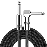
- [Low Noise High Fidelity] New bee guitar cord with 22AWG oxygen-free-copper center conductors provides enhanced signal transfer, the PVC insulation material and the high-density stranded copper double shield provide noise rejection for quiet operation
- [Ideal for Choice] New bee 1/4 straight and right angle metal connectors, ensures a secure connection in any instrument, pedal, or amp jack. L shape great fro on stage guitar stands
- [Wide Compatible] Suitable for mixer, power amplifier, microphone, effector, decoder, equalizer, electoronic organ, electric guitar, electric bass and other electronic equipments. Widely used in the installation of smart home, karaoke speaker, professional stage audio, etc
- [Easy Disassemble] Making incidental repairs quick and easy! Very nice, flexible cable
- [Dont Hesite to Order] 24 hours customer services and professional technology team are standing by
Tagged proteins can locate biosynthetic components
Instead of labeling a metabolite or cell wall, investigators can fuse a fluorescent tag to a selected enzyme or other biosynthetic protein. Fluorescence microscopy then shows where that tagged component appears: for example, whether it is diffuse, membrane-associated, concentrated at a pole or organized into visible foci.
This approach addresses a different question from MALDI imaging. It localizes a chosen protein rather than detecting the final antibiotic. Localization can suggest where a biosynthetic step occurs or whether several components occupy related regions, but it does not prove that the tagged protein is catalytically active there.
Tag placement and expression level matter. A fusion can alter folding, interactions, transport or activity, and overexpression can create artificial clusters. Useful controls include an untagged strain, a functional rescue test, expression at near-native levels and comparison with an independently labeled construct. A localization result becomes more persuasive when it agrees with biochemical activity, gene-perturbation data and the distribution of the product itself.
Windows Errors? Fix Them Before They Spread
Repair common Windows errors and clear accumulated junk for a smoother, more stable PC - no reinstall needed.Free scan · no reinstallOutdated Drivers Are Slowing You Down
One free scan finds every outdated or missing driver and matches the right update for your exact hardware.Free scan · exact hardware matchEmerging three-dimensional views of biosynthetic machinery
A 2024 conference abstract by Josyula and colleagues describes a developing program that combines cryo-focused ion beam milling and scanning electron microscopy (cryo-FIB-SEM) with cryo-electron tomography. These methods can reconstruct cellular ultrastructure in three dimensions. The same program describes multiphoton fluorescence microscopy and fluorescence-lifetime imaging microscopy (FLIM) to follow metabolites and tagged biosynthetic proteins.
In principle, correlative imaging could show how a biosynthetic megacomplex sits relative to membranes, vesicles or export structures while preserving information from both fluorescence and electron imaging. That would help address organizational questions that a two-dimensional fluorescence image cannot resolve.
The evidence currently supports describing this as an emerging research direction, not as a universally validated routine platform. Cryogenic preparation, correlation between imaging modalities and interpretation of large complexes each introduce technical constraints. Results still require biochemical and genetic validation.
Rank #4

- Digital Family Calendar: BSIMB digital calendar wall touch screen puts an end to your chaotic life. Designed for busy families, the digital calendar keeps you organised and in step with your family. Assign different colours and tasks to each family member to make family scheduling easier and more intuitive.
- Easy Setup and Auto-Sync: Plug in, connect to WLAN and auto-syn your electric calendar in minutes. Digital wall calendar compatible with Google calendar, iCloud calendar, Outlook calendar, Cozi calendar and Yahoo calendar. Add events directly on the device or through the ecalendar app. Electronic calendar stays current whether you're at work or traveling—no manual updates needed.
- All-In-One Home Assistant: Smart calendar builds habits and encourage independence with personalised chores. Display daily meal plan to reduce the daily ‘What's for dinner?’ stress. To-do lists keep track of everything you want. Turning chores into valuable achievements with stars and rewards, making life easier and more enjoyable.
- Manage Anytime Anywhere: BSIMB electronic calendar for family stay connected with the powerful ecalendar app. Access and manage your digital wall calendar anytime, anywhere with free features like stars & rewards, photo screensaver, chore charts, meals plans, to-do lists etc. Electronic planner paid features like Magic Import tailor advanced tools for users with special needs.
- Stay in Touch with Loved Ones: Share photos or videos from your phone to electronic wall calendar with app, instantly update your life moment, and keep in touch with your family, especially those who are far away; you can also surprise your smart calendar digital wall by sending him a wish card; it's also great for keeping tech-illiterate elders in touch with the youngsters.
How scientists build a convincing imaging study
- Define the biological question. Decide whether the goal is to map a product, a precursor, cell-wall construction, a target or a biosynthetic protein. This choice determines whether mass spectrometry, a probe, a protein tag or a correlative method is appropriate.
- Choose the sample and conditions. Record strain, growth stage, medium, interacting organisms and treatment. Spatial patterns can change with development and environment, so conditions are part of the result.
- Acquire the primary image. For MALDI imaging, collect molecular-ion maps; for fluorescence, collect probe or tagged-protein images with settings that allow comparison between samples.
- Run specificity and preparation controls. Include blank or matrix controls for mass spectrometry, unlabeled or competition controls for probes, and untagged or expression controls for protein fusions. Check for autofluorescence, quenching and probe-access limitations.
- Identify the chemical signal. Use tandem MS or another orthogonal chemical method before assigning a mass feature to a named antibiotic or intermediate.
- Connect signal to genotype. Use genome mining to find a plausible biosynthetic gene cluster, then test candidate genes with activation, deletion or other genetic perturbations.
- Test function. Compare isolates or extracts in antimicrobial activity assays and verify that changes in the image track changes in the compound or phenotype.
- Integrate the evidence. Report what the image establishes, what supporting data establish, and which steps remain inferential. Avoid presenting spatial correlation as proof of causation.
Method comparison
| Approach | Main readout | Best suited question | Key interpretive caution |
|---|---|---|---|
| MALDI imaging mass spectrometry | Spatial distribution of detectable molecular ions | Where do candidate metabolites occur in a colony or sample, and how do signals relate to neighboring organisms or conditions? | A mass feature needs chemical identification; spatial correlation does not prove the responsible genes or biological function. Tandem MS, genome mining and activity tests strengthen the assignment. |
| Fluorescent molecular probes, including fluorescent D-amino acids | Labeled cell-wall material or selected molecular targets | Where is cell-wall synthesis or a probe target detectable? | Probe specificity, envelope entry, background fluorescence, quenching and labeling effects can limit interpretation. |
| Tagged proteins and fluorescence imaging | Position or distribution of a labeled biosynthetic component | Where does a selected enzyme or protein appear? | The tag can perturb localization or activity; localization alone does not establish catalysis. |
| Cryo-electron and correlative imaging | Cellular ultrastructure and three-dimensional organization, potentially alongside fluorescence signals | How might biosynthetic complexes relate spatially to membranes and export structures? | The 2024 evidence is a conference abstract describing a research program, not a mature universal platform. |
There is no consistent head-to-head performance dataset across all of these approaches. The practical comparison therefore depends on the target, spatial scale, chemical specificity, sample preparation, live-cell compatibility and the orthogonal evidence available for validation.
Recommended Free Tools
Controls and common interpretation failures
Calling a mass peak a confirmed antibiotic
Mass-to-charge agreement narrows possibilities but may not distinguish isomers or adducts. Confirm identity with fragmentation and complementary chemistry before naming the compound.
Assuming colocalization proves interaction
Two signals in the same region may reflect shared transport, crowding or limited diffusion. Colocalization is a hypothesis-generating observation; biochemical interaction or genetic dependence requires separate tests.
Ignoring the cell envelope
A probe that cannot cross the envelope cannot report an intracellular target reliably. Uptake, efflux and permeability controls are essential when interpreting weak or uneven labeling.
Treating brightness as production rate
Fluorescence intensity and ion intensity are influenced by instrument settings, ionization, labeling efficiency and background. Quantitative claims require calibration and a validated relationship between signal and amount.
Free tools Windows power users keep installed
One-click scans. No signup required.
Overlooking tag-induced artifacts
A fluorescent fusion can mislocalize or impair an enzyme. Demonstrate that the tagged strain retains the relevant phenotype and compare it with an untagged control.
Confusing antibiotic production with antibiotic effects
Imaging where a drug accumulates in a susceptible bacterium, or where cell-wall damage appears after treatment, studies the drug’s action. Imaging a producing microbe’s metabolite or biosynthetic machinery studies production. The experiments may inform one another but answer different questions.
Can imaging show which genes produce an antibiotic?
Not on its own. Imaging can reveal where a candidate product or biosynthetic component occurs. Genome mining can identify a plausible gene cluster, and short-sequence-tagging peptidogenomics helped make that connection in the arylomycin study. To demonstrate causality, scientists generally perturb candidate genes and check whether the molecular image, isolated compound and antimicrobial activity change as predicted.
This division of labor is useful: imaging supplies spatial phenotype, mass spectrometry and chemistry supply molecular identity, and genetics supplies pathway attribution. A convincing claim requires agreement among those layers rather than a more dramatic image.
Choosing an approach for a new study
- Start with MALDI imaging when the unknown is a metabolite and its location in a colony, mixed culture or tissue is the main question.
- Use fluorescent D-amino acids when the focus is peptidoglycan synthesis, remodeling or cell-wall dynamics.
- Use a small-molecule fluorescent probe when a defined target is known and probe specificity, access and competition can be tested.
- Tag a protein when the position of a selected biosynthetic component matters more than direct detection of the product.
- Consider cryo-correlative methods for three-dimensional organization questions, recognizing their emerging status and demanding sample preparation.
Whichever method is chosen, plan the validating experiment at the same time as the imaging experiment. A map without chemical, genetic or functional follow-up is usually a useful lead, not a finished biosynthesis result.
Where the field is heading
The long-term goal is to connect chemistry, genetics and cell biology in the same specimen: identify a metabolite, locate it relative to its producer, place the responsible enzymes in a three-dimensional cellular context, and test its biological effect. Progress toward that goal is likely to come from correlative imaging, improved probes, more reliable chemical annotations and genome-mining strategies that expose previously silent pathways.
For now, the strongest studies remain deliberately multimodal. Molecular imaging answers “where,” while chemistry, genetics and microbiology establish “what,” “how” and “does it matter.”
Quick Recap
Product prices and availability are accurate as of the date/time indicated and are subject to change. Any price and availability information displayed on Amazon at the time of purchase will apply.
What’s actually slowing this PC down?
Pick the symptom - the matching free tool is one click away.

