Windows Errors? Fix Them Before They Spread
Repair common Windows errors and clear accumulated junk for a smoother, more stable PC - no reinstall needed.Free scan · no reinstallCrashes, No Sound, or Screen Glitches?
Random freezes, missing sound and display glitches usually trace back to one bad driver. Find and replace yours safely.Free scan · under a minuteScientists do not take a DNA photograph with one universal camera. They select an instrument that measures a particular signal—surface height, fluorescent blinking, or electron scattering—then prepare the DNA for that measurement and use analysis software to turn the signal into an interpretable image.
That distinction explains why DNA images can look radically different. An atomic-force microscope can trace a molecule attached to a surface; DNA-PAINT can reconstruct labeled strands from thousands of timed binding events; and a specialized electron microscope can record contrast from a single DNA fiber under tightly controlled conditions. Each picture answers a different structural question.

As an Amazon Associate I earn from qualifying purchases.
What a “DNA image” actually represents
A published image of DNA is usually a map of a physical signal, not a direct view comparable to a phone photograph. The signal may be the height of a probe moving over a surface, the position of fluorescent molecules that blink on and off, or the intensity of electrons transmitted through a specimen. Sample preparation is part of the measurement: attaching DNA to mica, adding fluorescent docking strands, or managing an electron beam can all influence what the final image means.
Interpretation therefore starts with the method. Scientists ask what was measured, what the preparation could have altered, and which features are supported by controls and analysis. A bright spot in a fluorescence reconstruction, for example, represents a localized emission event; a ridge in an AFM image represents topography relative to the substrate. Neither should automatically be read as a photograph of every atom in a double helix.
#1 Best Overall

- [Ultra-Lightweight Ear Buds Designed for Small Ears] Each earbud weighs only 3.7g and features a compact, ergonomic in-ear design made especially for small ears. Secure, low-profile, and comfortable for workouts, all-day wear or overnight sleeping.The ultra-slim profile sits flush in your ear with no hard edges poking or rubbing, so you can even sleep on your side without discomfort.
- [Immersive Stereo Sound with TOZO OrigX Technology] TOZO OrigX tuning delivers clear vocals, balanced mids, and natural stereo sound for music, podcasts, and videos.
- [Long Battery Life for Daily Use] Get up to 7 hours of playtime on a single charge, with up to 32 hours total using the charging case—ideal for workdays, commuting, and extended listening sessions.
- [Bluetooth 5.3 & Stable Connection] Bluetooth 5.3 provides fast pairing, stable wireless performance, and reduced dropouts as you move around home or office.
- [Deep Bass with Clear Vocals] High-performance drivers produce punchy bass while keeping vocals clean and detailed for everyday listening.
Atomic-force microscopy: tracing DNA’s surface shape
How the probe makes an image
Atomic-force microscopy (AFM) uses a very sharp probe mounted on a flexible cantilever. The probe scans across a sample while the instrument measures tiny changes in force or cantilever motion. Software converts those measurements into a topographic map: peaks, valleys and contours show the surface relief encountered by the probe.
For DNA experiments, molecules are generally immobilized on a flat substrate so the probe can follow them. AFM can reveal the shape and organization of surface-bound DNA, as well as protein–DNA complexes. Some protocols operate in liquid, allowing researchers to examine DNA in a more hydrated environment, but the molecule still interacts with the supporting surface. Surface chemistry is consequently part of the experiment rather than a neutral preliminary step.
Why mica and salt preparation matter
A 2019 JILA/NIST method used mica, a flat silicate mineral, with salt-based preparation to image DNA in liquid. According to NIST, the preparation extended DNA on the substrate so that eight times more of each molecule could be analyzed than with earlier preparation approaches. In the images described for that study, the measured DNA was about 680 nanometres long and about 2 nanometres high. Those dimensions are results from that preparation and experiment, not guaranteed values for every AFM sample.
Rank #2

- 【IP68 Waterproof Multifunctinal Smart Watch】Torjalph fitness activity tracker smart watch is IP68 waterproof technology ,you can wear it when swimming,handwashing,it's rain-proof which means that you perform multiple exercise modes outside without worrying about sweat or rain corroding your fitness smart watch,and this watch equipped with a 230 mAh large capacity battery magnetic charging ,and this sports activity tracker watch can be used 5-7 days If fully charged.
- 【Larger 1.83'' Full Screen Watch & Multi-dial Selection】1.83 inch full HD color touch screen smart watch for men / women provides excellent 240*284 resolution image quality ,This Bluetooth smart watch offers a better touch experience and remarkable immersion and ,more than 200 watch faces for you to choose from App"Da Fit ", you can change the dial style according to your mood to wear keeping the world on your wrist
- 【Accurate Activity Tracker Smart Watch 】sports watch activity tracker uses sweat and dust resistant breathable silicone strap, our Fitness Tracker Smart Watch for Android iOS Phones supports 12 sports modes,walking,running,bicycle,swimming,badminton,basketball,football,rope skipping. This bluetooth calls smart watch for men women is lightweight and stylish, bringing more convenient and relaxing sports experience while also providing more protection.
- 【Your 24 Hour Personal Daily Health Monitor】Fitness watch for women men full touch screen IP67 waterproof bluetooth smartwatch for Android iOS Phones with heart rate blood pressure monitor multifunction Andriod smart watch with steps counter sleep monitor sports fitness watch, monitoring blood oxygen saturation allows you to understand your physical condition in real time to track fitness progress ,to know your health status in time.
- 【More Functions Bluetooth Call Smart Watch】This fitness activity smart watch full screen for iPhone and Android phones includes a bunch of new features including:The bluetooth smart watch can hang up the phone, receive and display WhatsAPP /Text /Facebook /Twitter /Messenger reminder.Support sedentary reminder, alarm clock, stopwatch, timer,weather information,music control, remote camera control compatible with Android 5.0 and above or iOS 9.0 and above smartphones.
The same method was used to examine two protein–DNA complexes. By tuning how strongly DNA binds to the surface, researchers can seek a balance: attachment must be firm enough to scan, but not so disruptive that it hides the interaction being studied. JILA/NIST Fellow Tom Perkins described the aim this way: “We expect this new sample preparation method to pave the way to tuning the binding strength of DNA to a surface, which should facilitate studying the dynamics of protein-DNA complexes.”
A typical AFM workflow
- Prepare the substrate. Clean and condition mica or another suitable surface, then establish the salt and buffer conditions that control DNA adsorption.
- Introduce the DNA. Apply the DNA solution and allow molecules to attach and spread. Concentration, incubation time and ionic strength affect whether strands lie separately or form crowded regions.
- Rinse or exchange into imaging liquid. The chosen liquid environment removes unbound material while preserving the desired attachment.
- Scan with a calibrated probe. The probe follows the surface line by line. Excessive force can drag or deform strands; insufficient feedback can blur height information.
- Process the topography. Researchers flatten background slopes, identify individual contours and measure length, height, bends or protein-associated features. Processing must not create structures that were absent from the raw scan.
What AFM can and cannot establish
AFM is well suited to questions about molecular shape, arrangement and surface-associated interactions. It can show whether strands are extended, folded, clustered or associated with a protein under the chosen conditions. It does not automatically reveal the base sequence, and the need for a supporting surface means that an image may differ from DNA freely moving in solution. A representative conclusion must therefore include the substrate, buffer and attachment chemistry.
DNA-PAINT: building a super-resolution image from blinking molecules
The binding events behind the picture
DNA-PAINT is a fluorescence localization method rather than a direct topographic scan. Researchers attach short DNA “docking” strands to the structures they want to map. Dye-labeled complementary “imager” strands diffuse through the solution and bind briefly to those docking strands. Each binding event produces a fluorescent burst; when the imager strand leaves, the signal turns off.
Rank #3
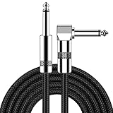
- [Low Noise High Fidelity] New bee guitar cord with 22AWG oxygen-free-copper center conductors provides enhanced signal transfer, the PVC insulation material and the high-density stranded copper double shield provide noise rejection for quiet operation
- [Ideal for Choice] New bee 1/4 straight and right angle metal connectors, ensures a secure connection in any instrument, pedal, or amp jack. L shape great fro on stage guitar stands
- [Wide Compatible] Suitable for mixer, power amplifier, microphone, effector, decoder, equalizer, electoronic organ, electric guitar, electric bass and other electronic equipments. Widely used in the installation of smart home, karaoke speaker, professional stage audio, etc
- [Easy Disassemble] Making incidental repairs quick and easy! Very nice, flexible cable
- [Dont Hesite to Order] 24 hours customer services and professional technology team are standing by
The microscope records thousands of frames containing these stochastic on-and-off events. Software estimates the position of each emitter from the light distribution in a frame, then combines the positions into a super-resolution reconstruction. The final image is therefore a statistical map assembled from many localized events, not a single exposure in which every label is simultaneously visible.
From sample preparation to molecule counting
The Nature Protocols workflow by Schnitzbauer, Strauss, Schlichthaerle, Schueder, Jungmann and colleagues covers the entire chain: sample preparation, multiplexed acquisition, simulation, image reconstruction, drift correction, quantitative qPAINT molecule counting and particle averaging. The authors identify Picasso as an integrated software package for these computational steps; Picasso is distributed through the Max Planck Institute of Biochemistry.
Developed DNA-PAINT approaches reported sub-5-nanometre spatial resolution and approximately 1-nanometre localization precision. These are technique-specific performance claims, not universal specifications for every microscope, label arrangement or sample. Resolution describes the ability to distinguish nearby features under defined conditions; localization precision describes how consistently the position of an individual event can be estimated. Neither number means that every base in an arbitrary DNA sample will be resolved.
Rank #4

- Digital Family Calendar: BSIMB digital calendar wall touch screen puts an end to your chaotic life. Designed for busy families, the digital calendar keeps you organised and in step with your family. Assign different colours and tasks to each family member to make family scheduling easier and more intuitive.
- Easy Setup and Auto-Sync: Plug in, connect to WLAN and auto-syn your electric calendar in minutes. Digital wall calendar compatible with Google calendar, iCloud calendar, Outlook calendar, Cozi calendar and Yahoo calendar. Add events directly on the device or through the ecalendar app. Electronic calendar stays current whether you're at work or traveling—no manual updates needed.
- All-In-One Home Assistant: Smart calendar builds habits and encourage independence with personalised chores. Display daily meal plan to reduce the daily ‘What's for dinner?’ stress. To-do lists keep track of everything you want. Turning chores into valuable achievements with stars and rewards, making life easier and more enjoyable.
- Manage Anytime Anywhere: BSIMB electronic calendar for family stay connected with the powerful ecalendar app. Access and manage your digital wall calendar anytime, anywhere with free features like stars & rewards, photo screensaver, chore charts, meals plans, to-do lists etc. Electronic planner paid features like Magic Import tailor advanced tools for users with special needs.
- Stay in Touch with Loved Ones: Share photos or videos from your phone to electronic wall calendar with app, instantly update your life moment, and keep in touch with your family, especially those who are far away; you can also surprise your smart calendar digital wall by sending him a wish card; it's also great for keeping tech-illiterate elders in touch with the youngsters.
A practical DNA-PAINT sequence
- Design docking sites. Place complementary DNA sequences at the molecular positions to be measured. Their spacing and accessibility determine which features can be localized.
- Attach labels and prepare the sample. Connect docking strands to the target structure and introduce dye-labeled imager strands in a controlled buffer.
- Acquire a time series. Record many frames so different docking sites produce temporally separated flashes. Multiplexed experiments can use different sequence pairs to image several targets.
- Localize emitters. Fit the fluorescence pattern from each event to estimate its center, reject poor fits and track signal quality over time.
- Correct drift and reconstruct. Mechanical or thermal drift can move the entire field during acquisition. Correction aligns the localizations before rendering the super-resolution image.
- Count and average when appropriate. qPAINT uses binding kinetics to estimate molecule numbers, while particle averaging can improve repeated structural patterns. These analyses depend on calibration and assumptions about binding behavior.
The published protocol describes completion in one to two days. That is the duration of the full procedure, including preparation and computational work, rather than a promise that a microscope will collect its data in that time.
How to read a DNA-PAINT reconstruction
A reconstructed dot or line indicates where labeled docking sites were localized. It is evidence about the arrangement of those sites and, with suitable calibration, their number. It is not an unlabeled image of the DNA backbone. Missing events can result from inaccessible docking strands or incomplete binding, while overlapping signals, drift and nonspecific interactions can create apparent distortions. Controls, simulations and repeated acquisitions help distinguish molecular organization from reconstruction artifacts.
Free tools Windows power users keep installed
One-click scans. No signup required.
Direct electron imaging: a specialized HRTEM demonstration
Electrons have much shorter wavelengths than visible light, but biological molecules present a difficult specimen. DNA contains relatively light atoms and therefore produces weak electron contrast; the beam can also damage or destroy the molecule. Researchers must balance dose, contrast, focus and specimen stability.
A 2015 Scientific Reports paper reported direct imaging of a single A-form double-stranded DNA fiber with spherical-aberration-corrected high-resolution transmission electron microscopy (HRTEM). The experiment relied on carefully selected preparation and imaging conditions. It demonstrates that direct structural detail is possible in a specialized setting, not that ordinary electron microscopy routinely photographs DNA bases or that every DNA sample can be imaged this way.
What the electron image means
In HRTEM, image intensity arises from how electrons scatter as they pass through the specimen and its support. Contrast depends on thickness, composition, focus and instrument settings. Radiation damage can alter the structure during exposure, so an apparent feature must be evaluated against dose history and specimen condition. The result is best treated as a carefully constrained structural measurement rather than a universal template for the double helix.
“DNA microscopy” can mean something else
The phrase DNA microscopy is also used for an optics-free technique that encodes physical images in DNA. A 2019 Nature Methods research highlight titled “Imaging with DNA” uses the term in that sense: DNA molecules store information about an image, and sequencing-based decoding reconstructs it. This is different from using AFM, fluorescence or electrons to image DNA molecules themselves.
How scientists decide what an image supports
Start with the measured signal
- AFM: surface topography measured by a scanning probe.
- DNA-PAINT: positions and timing of fluorescent binding events.
- HRTEM: electron-scattering contrast in a thin specimen.
Check preparation-specific effects
For AFM, ask how mica, salts and adsorption might have extended, compressed or oriented the strand. For DNA-PAINT, check whether labels and docking strands can reach every intended site and whether binding kinetics support counting. For HRTEM, examine specimen preparation, beam dose and evidence of radiation damage. A method can produce a sharp-looking image while still measuring only a subset of the molecule’s physical properties.
Separate observation from interpretation
An observed contour, localized spot or contrast feature is the instrument’s immediate output. Claims about a protein binding site, molecular count or helical form require calibration, controls and a model connecting that output to structure. Scientists compare images with simulations, known standards or repeated measurements and report uncertainty when preparation or signal quality limits the conclusion.
AFM, DNA-PAINT and HRTEM compared
| Method | Primary signal | Sample requirements | What the data can support | Main analysis |
|---|---|---|---|---|
| Atomic-force microscopy | Surface topography from probe–sample interactions | DNA attached to a surface; liquid protocols use substrates such as mica and controlled salt conditions | Shape, organization and surface-associated protein–DNA interactions | Topography correction, contour measurement and interpretation of surface effects |
| DNA-PAINT | Localized fluorescence from transient imager–docking-strand binding | Dye-labeled imager strands, complementary docking strands, controlled imaging buffer and long time-series acquisition | Super-resolution localization, multiplexed target maps and calibrated molecule counts | Emitter localization, drift correction, reconstruction, qPAINT counting and particle averaging |
| Spherical-aberration-corrected HRTEM | Electron-scattering contrast | Specialized specimen preparation, carefully chosen imaging conditions and dose management | Direct structural detail in a constrained single-fiber demonstration | Electron-image formation, focus and contrast assessment, plus radiation-damage evaluation |
Which approach answers which question?
- How is DNA arranged on a surface, or where does a protein sit on an immobilized strand? AFM is a natural choice when the surface and attachment chemistry can be controlled.
- Where are several molecular targets, and how many labeled sites are present? DNA-PAINT is useful when sequence-specific docking strands and quantitative localization are feasible.
- Can a carefully prepared individual DNA fiber produce direct electron contrast? HRTEM can address that specialized structural question, provided the experiment controls weak contrast and radiation damage.
- Is the phrase “DNA microscopy” referring to an encoded image rather than a molecular image? Check the method’s signal. Optics-free DNA encoding is a different technology from imaging DNA with a microscope.
The central lesson
Scientists “see” DNA by converting a physical interaction into data and then testing what that data can legitimately mean. AFM maps a surface-bound molecule, DNA-PAINT reconstructs positions from many fluorescent binding events, and HRTEM records electron contrast under exceptional conditions. None is a universal camera for the double helix. The most reliable interpretation always names the signal, the preparation, the analysis steps and the limits those choices impose.
Quick Recap
Product prices and availability are accurate as of the date/time indicated and are subject to change. Any price and availability information displayed on Amazon at the time of purchase will apply.
What’s actually slowing this PC down?
Pick the symptom - the matching free tool is one click away.

