Recommended Free Tools
MRI monitoring is a comparison process, not a verdict from one scan. A neuro-oncology team establishes an appropriate baseline, compares later images with it and with the patient’s prior scans, and then weighs measurable tumor changes alongside new lesions, neurologic status and corticosteroid use. The result helps classify disease as responding, stable or progressing, while accounting for treatment effects that can temporarily resemble growth.
The exact interpretation depends on tumor subtype, treatment, timing and the measurement method. RANO 2.0, a consensus framework for adult gliomas, standardizes much of this language, but its thresholds are clinical assessment tools—not an individual diagnosis or prognosis.

As an Amazon Associate I earn from qualifying purchases.
What serial MRI monitoring is designed to show
MRI produces detailed images of the brain, but treatment monitoring depends on how images change over time. Radiologists and neuro-oncologists compare a current scan with a defined reference scan, looking for changes in enhancing tumor, nonenhancing disease, edema and new lesions. They also review symptoms, neurologic examination findings and medication changes.
The Tool Desk
Outbyte PC Repair FREERepair Windows errors before they cause bigger problemsFix Now →Outbyte Driver Updater FREEFix the driver behind crashes, sound loss and screen glitchesFind Drivers →This longitudinal approach answers practical questions: Has measurable disease decreased, stayed within the range considered stable, or increased enough to raise concern for progression? Is a new area a genuine lesion, postoperative change, inflammation or a treatment effect? Has the patient’s clinical condition changed in a way that makes an imaging finding more urgent?
#1 Best Overall

- [Ultra-Lightweight Ear Buds Designed for Small Ears] Each earbud weighs only 3.7g and features a compact, ergonomic in-ear design made especially for small ears. Secure, low-profile, and comfortable for workouts, all-day wear or overnight sleeping.The ultra-slim profile sits flush in your ear with no hard edges poking or rubbing, so you can even sleep on your side without discomfort.
- [Immersive Stereo Sound with TOZO OrigX Technology] TOZO OrigX tuning delivers clear vocals, balanced mids, and natural stereo sound for music, podcasts, and videos.
- [Long Battery Life for Daily Use] Get up to 7 hours of playtime on a single charge, with up to 32 hours total using the charging case—ideal for workdays, commuting, and extended listening sessions.
- [Bluetooth 5.3 & Stable Connection] Bluetooth 5.3 provides fast pairing, stable wireless performance, and reduced dropouts as you move around home or office.
- [Deep Bass with Clear Vocals] High-performance drivers produce punchy bass while keeping vocals clean and detailed for everyday listening.
Why the comparison baseline matters
The reference scan is not always the first MRI after surgery. Under RANO 2.0, for a newly diagnosed adult glioma treated with radiotherapy, the post-radiotherapy MRI is used as the baseline for later response assessments. Surgery can leave irregular enhancement and other changes, so using the post-radiotherapy study creates a more consistent starting point for evaluating treatment.
For recurrent disease, the relevant comparison is generally the pretreatment scan obtained before the new therapy begins. A report that says “increased from baseline” therefore needs to be read with the date and purpose of that baseline in mind.
How clinicians read the images
Routine MRI sequences provide different types of evidence
A monitoring examination usually combines several sequences rather than relying on one picture. Contrast-enhanced images show areas where the blood–brain barrier is disrupted. T2-weighted and FLAIR images show nonenhancing abnormalities, including infiltrating tumor, edema and treatment-related change. Diffusion and perfusion sequences can add information about cellularity or blood flow, although they do not automatically settle the question of recurrence.
Free tools Windows power users keep installed
One-click scans. No signup required.
The American College of Radiology’s 2024 Brain Tumors guidance discusses MRI with intravenous contrast for post-treatment surveillance and emphasizes the difficulty of distinguishing recurrence, pseudoprogression and radiation necrosis. The contrast decision and protocol are made by the clinical team after considering the tumor, treatment and the patient’s medical history.
The tumor component being measured can differ
RANO 2.0 does not treat every glioma as an identical imaging problem. For IDH-wild-type glioblastoma, contrast-enhancing disease is the principal measurable component, with a specific exception when evaluating antiangiogenic treatment, where nonenhancing disease also matters. For IDH-mutated gliomas with a substantial nonenhancing component, a trial may assess both enhancing and nonenhancing portions.
Rank #2

- 【IP68 Waterproof Multifunctinal Smart Watch】Torjalph fitness activity tracker smart watch is IP68 waterproof technology ,you can wear it when swimming,handwashing,it's rain-proof which means that you perform multiple exercise modes outside without worrying about sweat or rain corroding your fitness smart watch,and this watch equipped with a 230 mAh large capacity battery magnetic charging ,and this sports activity tracker watch can be used 5-7 days If fully charged.
- 【Larger 1.83'' Full Screen Watch & Multi-dial Selection】1.83 inch full HD color touch screen smart watch for men / women provides excellent 240*284 resolution image quality ,This Bluetooth smart watch offers a better touch experience and remarkable immersion and ,more than 200 watch faces for you to choose from App"Da Fit ", you can change the dial style according to your mood to wear keeping the world on your wrist
- 【Accurate Activity Tracker Smart Watch 】sports watch activity tracker uses sweat and dust resistant breathable silicone strap, our Fitness Tracker Smart Watch for Android iOS Phones supports 12 sports modes,walking,running,bicycle,swimming,badminton,basketball,football,rope skipping. This bluetooth calls smart watch for men women is lightweight and stylish, bringing more convenient and relaxing sports experience while also providing more protection.
- 【Your 24 Hour Personal Daily Health Monitor】Fitness watch for women men full touch screen IP67 waterproof bluetooth smartwatch for Android iOS Phones with heart rate blood pressure monitor multifunction Andriod smart watch with steps counter sleep monitor sports fitness watch, monitoring blood oxygen saturation allows you to understand your physical condition in real time to track fitness progress ,to know your health status in time.
- 【More Functions Bluetooth Call Smart Watch】This fitness activity smart watch full screen for iPhone and Android phones includes a bunch of new features including:The bluetooth smart watch can hang up the phone, receive and display WhatsAPP /Text /Facebook /Twitter /Messenger reminder.Support sedentary reminder, alarm clock, stopwatch, timer,weather information,music control, remote camera control compatible with Android 5.0 and above or iOS 9.0 and above smartphones.
That is why a change in FLAIR signal, enhancement or edema cannot be interpreted in isolation. The tumor’s biology and the therapy being studied determine which component carries the most weight.
Two-dimensional and volumetric measurements
RANO 2.0 retains the maximum cross-sectional area as its primary measurement. In practice, the reader measures two perpendicular diameters on the most representative slice and calculates their product; the products from target lesions are then added. Volumetric measurement is an option and may represent three-dimensional change more directly, but the area and volume thresholds are different.
| Assessment choice | How it is used | Important qualification |
|---|---|---|
| Comparison scan | Post-radiotherapy MRI for a newly diagnosed adult glioma; pretreatment MRI for recurrent disease | The date and treatment context define which scan is the valid reference |
| Primary measurement | Maximum cross-sectional area, using products of perpendicular diameters | RANO 2.0 also permits volumetric assessment |
| Tumor component | Enhancing disease, nonenhancing T2/FLAIR disease, or both | The relevant component varies with tumor subtype and treatment |
| Clinical modifiers | New lesions, neurologic status and corticosteroid dose | A radiology percentage alone does not determine the response category |
What RANO 2.0 response categories mean
RANO 2.0 gives teams common thresholds for adult glioma assessments. The criteria are most useful when the reader knows which measurement method was used and what comparison point was selected.
| Category | RANO 2.0 measurement concept | Other conditions |
|---|---|---|
| Complete response | Disappearance of measurable lesions | The disappearance must be sustained for at least four weeks and accompanied by the applicable clinical and corticosteroid criteria |
| Partial response | At least a 50% decrease in the sum of products of perpendicular diameters, or at least a 65% reduction in volume | The improvement must persist for at least four weeks |
| Stable disease | Neither the response nor progression thresholds are met | Clinical status, steroid use and new lesions still matter |
| Progressive disease | At least a 25% increase in area, or at least a 40% increase in volume, measured against the specified baseline or the smallest prior measurement | A new measurable lesion may also qualify, subject to the confirmation rules applicable to the situation |
The “smallest prior measurement” is often called the nadir. Using the nadir prevents a tumor that initially shrank and then regrew from being judged only against its original size. Conversely, a modest change that does not reach a defined threshold may remain in the stable-disease category even when it deserves close clinical attention.
These thresholds are not promises about how a person will feel or how long treatment will work. They are a structured way to describe imaging and clinical data. A clinician may need to review the actual images, symptoms and medication record before deciding whether a treatment should change.
Rank #3
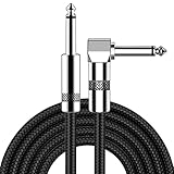
- [Low Noise High Fidelity] New bee guitar cord with 22AWG oxygen-free-copper center conductors provides enhanced signal transfer, the PVC insulation material and the high-density stranded copper double shield provide noise rejection for quiet operation
- [Ideal for Choice] New bee 1/4 straight and right angle metal connectors, ensures a secure connection in any instrument, pedal, or amp jack. L shape great fro on stage guitar stands
- [Wide Compatible] Suitable for mixer, power amplifier, microphone, effector, decoder, equalizer, electoronic organ, electric guitar, electric bass and other electronic equipments. Widely used in the installation of smart home, karaoke speaker, professional stage audio, etc
- [Easy Disassemble] Making incidental repairs quick and easy! Very nice, flexible cable
- [Dont Hesite to Order] 24 hours customer services and professional technology team are standing by
Why an MRI can look worse without proving tumor growth
Pseudoprogression after radiotherapy
Pseudoprogression is a treatment-related inflammatory or vascular change that resembles enlarging tumor on MRI. In glioblastoma during the first 12 weeks after radiotherapy and chemotherapy, published experience summarized in the 2023 RANO 2.0 update places its incidence at up to 30%–40%. This figure applies to that tumor and treatment window; it is not a rate for every brain tumor or every therapy.
What’s actually slowing this PC down?
Pick the symptom - the matching free tool is one click away.
For a clinically stable patient in this early period, RANO 2.0 recommends continuing treatment while confirming suspected progression with a repeat MRI or unequivocal pathology before labeling the scan as definite progression. The article gives four- or eight-week intervals as examples of confirmation timing in this specific context. They are examples, not a universal appointment schedule, and they should never be interpreted as a reason to delay urgent evaluation when neurologic function is worsening.
The time window is not identical for every tumor
In IDH-mutated gliomas and other glial tumors, pseudoprogression can persist beyond three months. A later scan that remains abnormal therefore still requires the tumor type, treatment and clinical course to be considered. The absence of a quick radiographic improvement does not, by itself, prove that treatment has failed.
Other treatment effects can mimic recurrence
Radiation necrosis, postoperative change, edema and inflammation can all produce enhancement or signal abnormalities. These possibilities overlap on conventional MRI. A report may describe “concerning for progression” rather than “definite progression” because the imaging appearance alone cannot separate these causes.
When advanced MRI is considered
Diffusion and perfusion
Diffusion MRI and dynamic susceptibility contrast perfusion MRI may provide additional clues. Restricted diffusion can reflect high cellularity but can also occur with treatment injury; perfusion measurements may suggest increased blood volume in active tumor but are affected by acquisition technique, leakage and therapy. RANO 2.0 states that these approaches need further validation before formal incorporation into its response criteria.
Rank #4

- Digital Family Calendar: BSIMB digital calendar wall touch screen puts an end to your chaotic life. Designed for busy families, the digital calendar keeps you organised and in step with your family. Assign different colours and tasks to each family member to make family scheduling easier and more intuitive.
- Easy Setup and Auto-Sync: Plug in, connect to WLAN and auto-syn your electric calendar in minutes. Digital wall calendar compatible with Google calendar, iCloud calendar, Outlook calendar, Cozi calendar and Yahoo calendar. Add events directly on the device or through the ecalendar app. Electronic calendar stays current whether you're at work or traveling—no manual updates needed.
- All-In-One Home Assistant: Smart calendar builds habits and encourage independence with personalised chores. Display daily meal plan to reduce the daily ‘What's for dinner?’ stress. To-do lists keep track of everything you want. Turning chores into valuable achievements with stars and rewards, making life easier and more enjoyable.
- Manage Anytime Anywhere: BSIMB electronic calendar for family stay connected with the powerful ecalendar app. Access and manage your digital wall calendar anytime, anywhere with free features like stars & rewards, photo screensaver, chore charts, meals plans, to-do lists etc. Electronic planner paid features like Magic Import tailor advanced tools for users with special needs.
- Stay in Touch with Loved Ones: Share photos or videos from your phone to electronic wall calendar with app, instantly update your life moment, and keep in touch with your family, especially those who are far away; you can also surprise your smart calendar digital wall by sending him a wish card; it's also great for keeping tech-illiterate elders in touch with the youngsters.
Advanced sequences are therefore complementary. They can help a multidisciplinary team decide whether a repeat scan, tissue sampling or a treatment discussion is warranted, but no single advanced parameter definitively diagnoses growth on its own.
Why conventional criteria remain important
Standardized measurements make scans comparable across institutions and clinical trials. They also provide a shared language when radiology, neuro-oncology, radiation oncology and neurosurgery review the same case. Advanced imaging can refine uncertainty, but it does not replace the baseline, the measured lesion, the clinical examination or the steroid history.
Independent reader supportYour contribution helps us test, update, and keep practical guides available for everyone.What happens during a monitoring cycle
- The team identifies the correct reference scan. The clinician confirms whether the case is newly diagnosed after radiotherapy or recurrent disease before choosing the comparison point.
- The MRI protocol is selected. The examination typically includes the sequences needed to evaluate enhancement and T2/FLAIR abnormality; contrast and optional advanced sequences depend on the clinical question.
- The radiologist compares current and prior images. Target lesions are measured using the agreed area or volumetric method, and new lesions or qualitative changes are recorded.
- The clinical record is reviewed. Neurologic symptoms, examination findings, corticosteroid dose and treatment dates are considered with the images.
- The team assigns or discusses a response category. If thresholds are met but pseudoprogression is plausible, the team may seek confirmation rather than immediately declaring treatment failure.
- The plan is individualized. The next scan, treatment decision or additional test depends on the tumor type, therapy, symptoms and degree of uncertainty; RANO criteria do not establish one schedule for every patient.
How to read an MRI report without overinterpreting it
A useful report review starts with context rather than the most alarming phrase. These questions help clarify what the radiologist and treating team mean:
- Which scan is being used as the baseline, and when was it performed?
- Is the reported change in contrast enhancement, T2/FLAIR abnormality, diffusion, perfusion or a combination?
- Was the change measured as a two-dimensional area or as a volume?
- Does the report identify a new measurable lesion?
- How long has it been since radiotherapy, surgery or the current drug treatment began?
- Could pseudoprogression, radiation necrosis, edema or postoperative change explain the appearance?
- Have symptoms, neurologic findings or corticosteroid dose changed?
- Is a confirmation MRI or another test being considered, and what clinical change would require earlier assessment?
Terms such as “stable,” “indeterminate” and “concerning for progression” describe different levels of certainty. They should be interpreted alongside the actual images and the treatment timeline, not converted into a percentage-based prediction.
Do these 3 things before closing this tab:
1Scan for outdated or missing drivers - takes under a minute2Clear out junk files and repair common Windows errors3Fix the driver behind crashes, sound loss and screen glitchesSituations that require extra caution
Early post-radiotherapy imaging
The first 12 weeks after glioblastoma radiotherapy are the setting in which pseudoprogression is especially common. A clinically stable patient with a concerning scan may be monitored for confirmation under the treating team’s plan. New weakness, seizures, confusion, severe headache or other neurologic deterioration changes the urgency and should be reported promptly rather than waiting for a routine comparison.
Immunotherapy
Immunotherapy can produce inflammatory imaging changes that complicate conventional response assessment. The iRANO framework was developed to address this problem. Its existence reinforces the broader point: treatment class and clinical status influence how an apparent early progression is interpreted.
Corticosteroid use
Steroids can reduce edema and improve symptoms while also affecting how response categories are assigned. RANO 2.0 includes corticosteroid dose among the clinical factors considered with imaging. A scan that appears improved while steroid requirements rise is not equivalent to an improvement accompanied by stable or declining steroid use.
What MRI monitoring can—and cannot—tell you
Serial MRI can show whether measurable disease has changed relative to a defined baseline, whether new lesions have appeared and whether the imaging pattern is compatible with response, stability or progression under a standardized framework. It can also reveal why a single scan may be ambiguous, particularly soon after radiotherapy.
PC Slower Than It Used to Be?
A free scan shows the junk files, broken settings and background clutter dragging Windows down - then fixes them in one click.Free scan · Windows 10 & 11Crashes, No Sound, or Screen Glitches?
Random freezes, missing sound and display glitches usually trace back to one bad driver. Find and replace yours safely.Free scan · under a minuteIt cannot independently determine the cause of every enhancement, replace a neurologic examination or provide an individual prognosis from one percentage. The most reliable interpretation combines the tumor subtype, treatment dates, comparison scan, measurement method, symptoms, steroid use and—when necessary—confirmatory imaging or pathology. Those safeguards are what make MRI monitoring clinically useful rather than merely a sequence of pictures.
Quick Recap
Product prices and availability are accurate as of the date/time indicated and are subject to change. Any price and availability information displayed on Amazon at the time of purchase will apply.

