The Tool Desk
Outbyte PC Repair FREEClear out junk files and repair common Windows errorsFree Scan →Outbyte Driver Updater FREEScan for outdated or missing drivers - takes under a minuteDriver Scan →An unruptured brain aneurysm does not automatically need an operation. A neurovascular team balances the aneurysm’s estimated chance of rupture against the risks of clipping, coiling, flow diversion, or continued observation, while considering your age, health, anatomy, family history and preferences. Some aneurysms are followed with repeat imaging; others merit treatment because they are growing, symptomatic, large, irregularly shaped, high-risk in location, or associated with important personal or family factors.
The practical plan is individualized. There is no safe, universal scan interval or single procedure that suits every aneurysm. Imaging and clinical review show whether the aneurysm is stable, changing or causing symptoms, and a specialist team uses that information to recommend surveillance or intervention.
As an Amazon Associate I earn from qualifying purchases.
What an unruptured brain aneurysm is—and why management varies
A cerebral (brain) aneurysm is a weakened area in an artery that bulges outward. “Unruptured” means it has not bled into the space around the brain. Many are discovered incidentally during a scan for another problem. The National Institute of Neurological Disorders and Stroke (NINDS) emphasizes that “not all cerebral aneurysms need treatment.” (NINDS patient information.)
Observation is not the same as ignoring an aneurysm. It usually means a documented imaging plan, clinical review and active management of factors such as high blood pressure and smoking. Intervention is considered when the expected harm from a future rupture is judged greater than the immediate risks of a procedure. A small aneurysm may still deserve treatment if it is enlarging, irregular, symptomatic or in a higher-risk location; a larger aneurysm may sometimes be observed when intervention would be especially hazardous.
#1 Best Overall
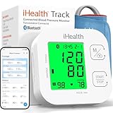
- Clinically Accurate: Easy Operation by two buttons, Advanced Accuracy, No Calibration required.
- Large Multi-Color Backlit Display: The large, oversized numbers make reading the results from this upper arm blood pressure monitoring device a breeze. Coded (GREEN/YELLOW/RED) display tells if your blood pressure readings are optimal or not.
- Easy Management: Manage and track up to 99 readings on your blood pressure monitor and unlimited readings on your smartphone with iHealth's free iOS and Android applications ( iOS 12.0 or later. Android 9.0 or later.Requires Bluetooth 4.0.and up).
- Heart Rhythm Disturbances: This unit automatically identifies and alerts you to any heart rhythm disturbances. If detected, a warning symbol will be displayed after the measurement is taken.
- What You Get: 1 x blood pressure monitor that meets ESH 2010 performance standards, 4 AAA batteries, 1 wide range cuff that fits upper arms with Circumference of 8.7"-16.5" (22-42cm),1 Instruction manual, our worry-free 12-month warranty and friendly California-based customer service.
These decisions are different from the emergency management of a ruptured aneurysm. A sudden bleed requires urgent hospital treatment, whereas an incidental unruptured aneurysm allows time for specialist assessment and shared decision-making.
How aneurysms are found and characterized
CT angiography (CTA)
CTA combines computed tomography with injected contrast to show arteries and an aneurysm’s relationship to surrounding vessels and bone. It is widely available and fast, which is useful when a clinician needs a detailed vascular picture promptly. Radiation exposure and contrast-related considerations are reviewed against the diagnostic benefit.
MR angiography (MRA)
MRA uses magnetic resonance techniques to display blood vessels. Depending on the technique, it may be performed with or without contrast and does not use ionizing radiation. Motion, implanted devices, kidney function and local imaging protocols can affect whether it is suitable. For following an unruptured aneurysm, the AHA/ASA summary identifies CT and MR angiography as the most sensitive noninvasive methods (AHA/ASA summary).
Catheter cerebral angiography
In catheter angiography, an interventional specialist guides a thin tube through an artery, usually from the wrist or groin, injects contrast and records detailed X-ray images. It can define the exact neck, branches, size and shape of an aneurysm when noninvasive scans leave uncertainty or when a treatment is being planned. Because it is invasive, the team weighs its information against risks such as bleeding, vessel injury or stroke.
Rank #2

- One-Button Design: This Oklar blood pressure monitor features a one-button operation, making measurement easy and convenient; with just a touch, you can quickly take a measurement and get an accurate reading
- Large LED Display: The backlit LED screen with clear digits makes it easy for anyone to read quickly; you can get readings within 1 minute, and it's user-friendly for the elderly
- Stores Up to 240 Readings: Supports two users, storing 120 readings per user; this feature makes it easy to track and monitor blood pressure trends for you and your family over time
- Convenient Dual Power Supply: Allows to operate by using either 3AA batteries or a Type-C interface (Notice: Use the provided Type-C cable if no batteries are installed; This product is not rechargeable) ; its compact size and dual supply capabilities ensure it's ready for use at home or on the go
- Adjustable Arm Cuff: The arm cuff range from 8.7 - 15.7 inches (22-40 cm) fits your size, ensuring a comfortable and secure fit for most adults
What the first scan report should establish
- Whether the finding is a true aneurysm or another vascular shape.
- Its maximum dimensions and whether the contour is smooth or has a daughter sac, lobulation or other irregularity.
- Its artery and precise location, including whether important branch vessels arise nearby.
- Whether there are additional aneurysms or signs of prior bleeding.
- Which imaging method will provide the most reliable baseline for later comparison.
What monitoring involves
Monitoring generally combines repeat vascular imaging with a clinical appointment. The clinician compares each study with the original images, not only the written report, because small measurement differences can reflect scan technique or angle. Ask how the team will define meaningful growth and whom to contact if symptoms change.
There is no one follow-up interval that is safe for every patient. The timing depends on aneurysm anatomy, confidence in the baseline measurement, age and health, treatment risk, other aneurysms and local practice. NICE guidance, written for the UK, says follow-up decisions should account for the original intervention, other aneurysms, estimated risk of further bleeding, risks of additional investigation or intervention and the person’s preference (NICE NG228 recommendations). That principle also illustrates why an internet-generated schedule should not replace your specialist’s plan.
A typical surveillance process
- Confirm the baseline. A neurovascular specialist reviews CTA, MRA and, when needed, catheter angiography to establish the most accurate size, shape and location.
- Set a personalized review date. The team records when the next scan and appointment should occur, taking account of image quality, estimated risk and your health.
- Use the same or a deliberately chosen method. Keeping technique and measurement conventions consistent improves the ability to detect a real change.
- Compare images and symptoms. Growth, new neurological symptoms, headaches with concerning features or compression of nearby nerves can change the recommendation.
- Reconsider the plan at each review. Stable imaging may support continued observation; documented enlargement or a new high-risk feature may lead to a treatment discussion.
The AHA/ASA 2015 guideline summary states that aneurysms with documented enlargement should be offered treatment when there are no comorbidities that make treatment unsuitable (AHA/ASA guideline summary). This is guideline context, not an automatic command: current specialists still weigh the individual’s anatomy, health and treatment options.
Recommended Free Tools
How clinicians estimate rupture and treatment risk
A multidisciplinary team—typically including a neurosurgeon and an interventional neuroradiologist—should review a non-ruptured aneurysm. NICE recommends discussing the plan and alternatives after considering size, location, estimated lifetime rupture risk, the estimated risk of each treatment, coexisting illnesses and the person’s preferences (NICE recommendations). NINDS also lists age, overall health, family and medical history, and aneurysm characteristics as relevant factors (NINDS).
Rank #3

- CONFIDENCE IN EVERY READING: Monitor your heart health with the OMRON Bronze Upper Arm Blood Pressure Monitor's Advanced Accuracy Technology; Store up to 14 readings to track your progress
- SAFEGUARD YOUR HEART: Detect heartbeat irregularities during routine blood pressure measurements with OMRON's Irregular Heart Beat Detection, helping you and your doctor determine if treatment or further evaluation is needed
- START YOUR DAY WITH CONFIDENCE: Know when your blood pressure is elevated with Hypertension Indicator alerts; Regular monitoring can reveal irregular fluctuations before they lead to serious complications
- TURN NUMBERS INTO INSIGHTS: Connect effortlessly to the free OMRON connect app via Bluetooth to store unlimited blood pressure readings, track trends, and share reports with your doctor; Compatible with Apple Health and Google Fit
- READY OUT OF THE BOX: Includes everything you need to start monitoring your heart health; OMRON monitor, wide-range D-ring blood pressure cuff (fits arms 9"– 17"), storage case, instruction manual, quick start guide, and four AA batteries
Size is important but is not a stand-alone verdict. Location changes the consequences of rupture and the technical difficulty of treatment. Shape matters because an irregular or lobulated sac can behave differently from a smooth one. Growth on reliable side-by-side imaging is a particularly important warning sign. A previous subarachnoid hemorrhage, multiple aneurysms, a strong family history, uncontrolled hypertension, smoking and certain inherited conditions may also affect the discussion. Your clinicians may use a risk score, but the number is an aid to conversation rather than a guarantee for one person.
The team also estimates procedural risk: access to the aneurysm, nearby branch arteries, anesthesia and surgical risk, age-related recovery, kidney or heart disease, blood-thinning medicines and the consequences of a possible stroke. AHA/ASA advises that treatment be performed at high-volume centers experienced in aneurysm care (AHA/ASA summary).
Treatment approaches
Clipping, coiling and flow diversion are specialist procedures, not consumer treatments. The choice depends on the aneurysm’s neck, branch vessels, size, shape and location, as well as your health and preferences. In some cases, more than one approach is technically possible; in others, observation is safer.
Do these 3 things before closing this tab:
1Fix the driver behind crashes, sound loss and screen glitches2Clear out junk files and repair common Windows errors3Scan for outdated or missing drivers - takes under a minute| Approach | How it works | Potential advantages | Important limitations and follow-up |
|---|---|---|---|
| Microsurgical clipping | Through an operation, the neurosurgeon places a small clip across the aneurysm neck so blood cannot enter the sac. | Can provide durable exclusion when the anatomy is favorable. NINDS says aneurysms that are completely clipped generally do not recur. | Requires open surgery, anesthesia and recovery. Risks include stroke, bleeding, infection, seizures or injury to nearby brain structures; the individual risk depends on anatomy and health. |
| Endovascular coiling | A catheter is advanced through an artery and detachable platinum coils are packed into the aneurysm to reduce blood flow and encourage clotting. | Less invasive than open clipping and may suit aneurysms that are difficult to reach surgically. | Coils can compact or the aneurysm can reopen, so some patients need additional imaging and another procedure. Branch-vessel anatomy and the need for blood-thinning medicines are considered. |
| Flow diversion | A stent-like device is placed in the parent artery to redirect blood away from the aneurysm, allowing the sac to seal over time. | Can be useful for some very large aneurysms or those not amenable to clipping or coiling, as described by NINDS. | Requires suitable parent-artery anatomy and antiplatelet medication. It is not appropriate for every location or patient and carries risks such as clotting, bleeding or blockage of branch vessels. |
Each option has procedure-specific risks and may require follow-up imaging. A recommendation should explain why the proposed method fits your anatomy, what alternatives were considered and what would happen if the aneurysm recurs or remains partly filled.
Rank #4

- Quick & Accurate Readings: Get reliable blood pressure measurements in under a minute with one-touch operation, perfect for easy daily monitoring
- Large LCD Display & Dual User Mode: Features a clear, easy-to-read screen; Store and track readings separately for two users (120 memories each), suitable for family use
- Adjustable Wide-Range Cuff: The comfortable cuff fits arms 22-40cm (8.7"-15.7")
- Clear Time Display: Automatically records time and date with each reading for easy trend tracking
- Convenient Power & Complete Kit: Use the included Type-C power cable or 3xAA batteries (not included); Comes ready with cuff, storage bag, power cord, quick guide, and manual
What to expect in a treatment discussion
Before the procedure
- Review every scan, including the original images and any earlier studies.
- Discuss anesthesia, hospital stay, likely recovery, medication changes and restrictions.
- Identify medicines that affect bleeding, allergies to contrast, kidney disease and other conditions.
- Clarify whether antiplatelet or anticoagulant medication is required and for how long.
- Ask how the team will monitor for complications and when the first post-treatment scan will occur.
After clipping
Recovery follows an operation and may involve an intensive-care observation period, wound care and gradual return to activity. Imaging may be used to confirm complete occlusion or to assess another aneurysm. A completely clipped aneurysm generally does not recur, but that does not eliminate the need to address other aneurysms or vascular risk factors.
After coiling or flow diversion
Endovascular treatment usually involves a shorter access-site recovery, but the aneurysm and treated artery still need surveillance. Coiled aneurysms can recur, and flow-diverter patients must follow the antiplatelet plan precisely. Do not stop prescribed medication without the treating team’s instruction.
Independent reader supportYour contribution helps us test, update, and keep practical guides available for everyone.Reducing modifiable risks
NINDS advises people with unruptured aneurysms to address high blood pressure aggressively and make a plan to stop smoking or using drugs such as cocaine (NINDS guidance). AHA/ASA identifies smoking and hypertension as modifiable factors associated with unruptured aneurysm cohorts (AHA/ASA summary).
Work with a clinician on blood-pressure targets, prescribed medicines, activity, sleep and a smoking-cessation plan. A home blood-pressure monitor can be an optional way to record readings for that conversation; the cited guidance does not establish that any particular device treats an aneurysm or prevents rupture. Lifestyle changes lower vascular risk but do not make rupture risk zero.
Best Value

- CONFIDENCE IN EVERY READING: The OMRON Iron Upper Arm Blood Pressure Monitor's Advanced Accuracy Technology helps you monitor your heart health
- SAFEGUARD YOUR HEART: OMRON's Irregular Heart Beat Detection identifies heartbeat irregularities during routine blood pressure measurements, helping you and your doctor determine if treatment or further evaluation is needed
- START YOUR DAY WITH CONFIDENCE: Count on quick and accurate readings with simple one-touch operation; Just press one button; Regular monitoring can reveal irregular fluctuations before they lead to serious complications
- MORE ACCURATE RESULTS: Body Movement Detection alerts help enhance the accuracy of your blood pressure readings
- READY OUT OF THE BOX: Includes everything you need for your blood pressure kit; OMRON monitor, wide-range D-ring blood pressure cuff (fits arms 9"-17"), instruction manual, and quick start guide
Emergency symptoms: when to call for help
Call your local emergency number immediately for a sudden, severe “thunderclap” headache, especially if it reaches maximum intensity within seconds. Emergency symptoms can also include weakness or numbness on one side, trouble speaking or understanding, loss or blurring of vision, collapse, seizure, confusion, vomiting or a stiff neck. These signs can indicate bleeding or another stroke emergency even if you have never been told that your aneurysm is at high risk.
Do not drive yourself. NHS guidance specifically tells people with possible brain-aneurysm emergency symptoms not to drive to emergency care (NHS brain aneurysm guidance). Use local emergency services; access instructions differ by country.
Questions to take to a neurovascular specialist
- What are the aneurysm’s exact size, location, shape and neck anatomy?
- Has it changed compared with every prior scan, and how certain is that measurement?
- What is my estimated lifetime rupture risk, and which assumptions affect it?
- What is the estimated risk of clipping, coiling, flow diversion or another option for me?
- Why is observation safer than treatment—or why does treatment now offer more benefit?
- Which specialists will review my case, and is this a high-volume aneurysm center?
- What imaging method and timing do you recommend, and who will track the appointment?
- Would treatment require antiplatelet or anticoagulant medication?
- What symptoms should prompt an urgent call before the next appointment?
- Should relatives be assessed because of my family history or an inherited condition?
Monitoring after a ruptured aneurysm is different
If an aneurysm has already bled, the immediate goal is to prevent rebleeding and protect the brain from complications; treatment is urgent and may involve endovascular or surgical expertise. The 2023 AHA/ASA guideline for aneurysmal subarachnoid hemorrhage recommends specialist evaluation of treatment risks and benefits according to patient and aneurysm characteristics (2023 AHA/ASA guideline). Follow-up after that episode can include checking the treated aneurysm, looking for additional aneurysms and assessing the risk of further bleeding. NICE’s factors for choosing follow-up imaging—initial treatment, other aneurysms, further-bleeding risk, investigation and intervention risks, and patient preference—apply in that context (NICE NG228).
What’s actually slowing this PC down?
Pick the symptom - the matching free tool is one click away.
For an incidental, unruptured aneurysm, the central decision remains whether surveillance or preventive treatment offers the better balance for that individual. Keep the written imaging plan, report new neurological symptoms promptly and attend every specialist review.
Quick Recap
Product prices and availability are accurate as of the date/time indicated and are subject to change. Any price and availability information displayed on Amazon at the time of purchase will apply.

