Recommended Free Tools
Yes. X-ray microscopy can damage a specimen when absorbed photons deposit enough energy to cause radiolysis, break chemical bonds, or change its structure and chemistry. The risk is not described by one universal “safe dose”: it depends on the material, hydration, thickness, temperature, photon energy, imaging mode, feature being measured, and the endpoint you use to define damage. A picture can remain interpretable while the specimen’s molecular state has already changed.
What “damage” means in X-ray microscopy
Damage is any radiation-induced change that compromises the experiment’s purpose. In a biological sample, that might mean broken disulfide bridges, altered molecular structure, mass loss, bubbling, or chemical-state changes. In an elemental-fluorescence experiment, the fluorescence signal may still be measurable even after the molecular structure has been altered. Conversely, a small morphological change may matter more than a modest loss of signal in an imaging study. You therefore need to state the damage endpoint rather than treating “damage” as a single yes-or-no property.

As an Amazon Associate I earn from qualifying purchases.
Absorbed X-rays transfer energy to the specimen. That energy can produce ionization and secondary electrons, initiating radiolysis and chemical reactions. Ray F. Egerton and Colin Nave describe radiolysis as “a major problem in X-ray microscopy” as brighter synchrotron beams and tighter focusing deliver more photons to smaller volumes. The same concentrated beam that improves signal can accelerate the processes that change the specimen.
The Tool Desk
Outbyte PC Repair FREERepair Windows errors before they cause bigger problemsFix Now →Outbyte Driver Updater FREEFix the driver behind crashes, sound loss and screen glitchesFind Drivers →Why dose and resolution are linked
Dose is the energy deposited per unit mass of specimen, measured in grays (Gy). Image quality, however, is governed by whether enough detected photons distinguish a feature from its surroundings despite shot noise. As the target feature becomes smaller, the feature generally contributes less signal, so more fluence and absorbed dose are needed to reach a chosen signal-to-noise ratio. If the specimen changes before that exposure is complete, the experiment has reached a damage-limited resolution.
#1 Best Overall

- [Ultra-Lightweight Ear Buds Designed for Small Ears] Each earbud weighs only 3.7g and features a compact, ergonomic in-ear design made especially for small ears. Secure, low-profile, and comfortable for workouts, all-day wear or overnight sleeping.The ultra-slim profile sits flush in your ear with no hard edges poking or rubbing, so you can even sleep on your side without discomfort.
- [Immersive Stereo Sound with TOZO OrigX Technology] TOZO OrigX tuning delivers clear vocals, balanced mids, and natural stereo sound for music, podcasts, and videos.
- [Long Battery Life for Daily Use] Get up to 7 hours of playtime on a single charge, with up to 32 hours total using the charging case—ideal for workdays, commuting, and extended listening sessions.
- [Bluetooth 5.3 & Stable Connection] Bluetooth 5.3 provides fast pairing, stable wireless performance, and reduced dropouts as you move around home or office.
- [Deep Bass with Clear Vocals] High-performance drivers produce punchy bass while keeping vocals clean and detailed for everyday listening.
The 1/d4 relationship is a model, not a rule for every microscope
An International Union of Crystallography review published in 2020 describes approximate 1/d4 scaling of required dose and fluence with target resolution d for uniform-density voxels. Halving the target voxel size therefore increases the modeled requirement by roughly a factor of 16 under those assumptions. The relationship is useful for understanding why extreme resolution becomes expensive in dose, but it is not a universal conversion table.
Contrast mechanism, object thickness, attenuation, feature geometry, reconstruction efficiency, background, and the model used for radiation damage can all change the result. A thin, strongly absorbing feature and a weak-contrast protein feature in water do not incur the same dose penalty at the same nominal resolution. Report the resolution definition and contrast criterion whenever you compare experiments.
Published resolution figures are conditional
| Figure | What it describes | Conditions and qualification |
|---|---|---|
| 3 nm | Most optimistic theoretical estimate cited for lensless water-window imaging of protein in water or ice | From the 2020 IUCr review; depends on its stated assumptions and lacks sufficient observational evidence to select a definitive damage model for frozen-hydrated biological material. |
| About 10 nm | Estimated resolution limit for frozen-hydrated biological objects with natural protein–water contrast at Rose-criterion image quality | Howells and colleagues, 2009; a historical theoretical estimate, not a guarantee for every current microscope or specimen. |
| 8 nm absorption; 6 nm phase | Modeled damage-limited resolution for a protein feature in ice at 500 eV | Egerton and Nave, 2026, using the stated calculation in which dose equals 100 times the DLR value. |
| 14 nm absorption; 20 nm phase | Modeled values for the corresponding 8 keV case | Egerton and Nave, 2026; comparative model outputs, not universal performance limits. |
The 2026 calculations also caution that real instruments may be limited by optics before the specimen reaches the modeled damage limit. A microscope that cannot form or detect a 6 nm feature gains nothing by exposing a sample long enough to reach a theoretical 6 nm damage threshold.
PC Slower Than It Used to Be?
A free scan shows the junk files, broken settings and background clutter dragging Windows down - then fixes them in one click.Free scan · Windows 10 & 11Crashes, No Sound, or Screen Glitches?
Random freezes, missing sound and display glitches usually trace back to one bad driver. Find and replace yours safely.Free scan · under a minuteRank #2

- 【IP68 Waterproof Multifunctinal Smart Watch】Torjalph fitness activity tracker smart watch is IP68 waterproof technology ,you can wear it when swimming,handwashing,it's rain-proof which means that you perform multiple exercise modes outside without worrying about sweat or rain corroding your fitness smart watch,and this watch equipped with a 230 mAh large capacity battery magnetic charging ,and this sports activity tracker watch can be used 5-7 days If fully charged.
- 【Larger 1.83'' Full Screen Watch & Multi-dial Selection】1.83 inch full HD color touch screen smart watch for men / women provides excellent 240*284 resolution image quality ,This Bluetooth smart watch offers a better touch experience and remarkable immersion and ,more than 200 watch faces for you to choose from App"Da Fit ", you can change the dial style according to your mood to wear keeping the world on your wrist
- 【Accurate Activity Tracker Smart Watch 】sports watch activity tracker uses sweat and dust resistant breathable silicone strap, our Fitness Tracker Smart Watch for Android iOS Phones supports 12 sports modes,walking,running,bicycle,swimming,badminton,basketball,football,rope skipping. This bluetooth calls smart watch for men women is lightweight and stylish, bringing more convenient and relaxing sports experience while also providing more protection.
- 【Your 24 Hour Personal Daily Health Monitor】Fitness watch for women men full touch screen IP67 waterproof bluetooth smartwatch for Android iOS Phones with heart rate blood pressure monitor multifunction Andriod smart watch with steps counter sleep monitor sports fitness watch, monitoring blood oxygen saturation allows you to understand your physical condition in real time to track fitness progress ,to know your health status in time.
- 【More Functions Bluetooth Call Smart Watch】This fitness activity smart watch full screen for iPhone and Android phones includes a bunch of new features including:The bluetooth smart watch can hang up the phone, receive and display WhatsAPP /Text /Facebook /Twitter /Messenger reminder.Support sedentary reminder, alarm clock, stopwatch, timer,weather information,music control, remote camera control compatible with Android 5.0 and above or iOS 9.0 and above smartphones.
What dose can a sample tolerate?
There is no single tolerance value for “an X-ray microscopy sample.” A meaningful dose number must be paired with the specimen, preparation, beam conditions, and criterion used to call the sample damaged. A dose that preserves a broad elemental map may destroy a fragile molecular structure; a dose that changes morphology may leave elemental identity intact.
For scale, a 2014 IUCr review estimated approximately 1300 MGy for a particular two-dimensional fluorescence scan of a protein-dominated sample using 10 keV X-rays, with the review’s stated flux, pixel size, and dwell time. That is an example calculation for that geometry and acquisition, not a permissible threshold for proteins generally. It should not be transferred to a different thickness, energy, scan strategy, or endpoint.
When a paper reports a dose, check whether it is incident or absorbed, total or per projection, averaged over the specimen or concentrated in a focal volume, and whether it is measured or modeled. Also check whether the number is tied to loss of diffraction quality, a chemical change, a visible structural alteration, a signal-to-noise target, or another criterion. Without those qualifiers, two dose values can look comparable while describing different physical outcomes.
Rank #3
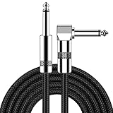
- [Low Noise High Fidelity] New bee guitar cord with 22AWG oxygen-free-copper center conductors provides enhanced signal transfer, the PVC insulation material and the high-density stranded copper double shield provide noise rejection for quiet operation
- [Ideal for Choice] New bee 1/4 straight and right angle metal connectors, ensures a secure connection in any instrument, pedal, or amp jack. L shape great fro on stage guitar stands
- [Wide Compatible] Suitable for mixer, power amplifier, microphone, effector, decoder, equalizer, electoronic organ, electric guitar, electric bass and other electronic equipments. Widely used in the installation of smart home, karaoke speaker, professional stage audio, etc
- [Easy Disassemble] Making incidental repairs quick and easy! Very nice, flexible cable
- [Dont Hesite to Order] 24 hours customer services and professional technology team are standing by
Does cryogenic cooling prevent X-ray damage?
No. Cooling can slow or redirect some damage pathways, but a cryogenic specimen continues to accumulate radiation damage. Freezing also changes diffusion, radical mobility, and sometimes the chemistry being measured; it does not make the sample dose-proof.
What’s actually slowing this PC down?
Pick the symptom - the matching free tool is one click away.
A 2010 study of specific protein crystals compared 50 K with 100 K. Using the study’s crystallographic R-free criteria, elastase tolerated 2.6 times the dose at 50 K, while cubic insulin tolerated 3.9 times the dose. Those are results for those crystals and that structural endpoint. They are not universal multipliers for cells, tissues, hydrated polymers, or fluorescence and phase-contrast measurements.
Use cryogenic preparation when it fits the specimen and the measurement, but still set a dose budget. Confirm that the cryogenic state preserves the feature of interest, and distinguish improved structural survival from preservation of chemical state or elemental signal.
Rank #4

- Digital Family Calendar: BSIMB digital calendar wall touch screen puts an end to your chaotic life. Designed for busy families, the digital calendar keeps you organised and in step with your family. Assign different colours and tasks to each family member to make family scheduling easier and more intuitive.
- Easy Setup and Auto-Sync: Plug in, connect to WLAN and auto-syn your electric calendar in minutes. Digital wall calendar compatible with Google calendar, iCloud calendar, Outlook calendar, Cozi calendar and Yahoo calendar. Add events directly on the device or through the ecalendar app. Electronic calendar stays current whether you're at work or traveling—no manual updates needed.
- All-In-One Home Assistant: Smart calendar builds habits and encourage independence with personalised chores. Display daily meal plan to reduce the daily ‘What's for dinner?’ stress. To-do lists keep track of everything you want. Turning chores into valuable achievements with stars and rewards, making life easier and more enjoyable.
- Manage Anytime Anywhere: BSIMB electronic calendar for family stay connected with the powerful ecalendar app. Access and manage your digital wall calendar anytime, anywhere with free features like stars & rewards, photo screensaver, chore charts, meals plans, to-do lists etc. Electronic planner paid features like Magic Import tailor advanced tools for users with special needs.
- Stay in Touch with Loved Ones: Share photos or videos from your phone to electronic wall calendar with app, instantly update your life moment, and keep in touch with your family, especially those who are far away; you can also surprise your smart calendar digital wall by sending him a wish card; it's also great for keeping tech-illiterate elders in touch with the youngsters.
How sample and experiment design change the risk
- Material and hydration: Organic, aqueous, frozen-hydrated, crystalline, and inorganic specimens have different absorption and radiolysis pathways.
- Thickness and depth: Absorption through a thick object changes both the photons reaching a feature and the dose deposited along the path. A surface feature and an equally small feature deep inside the specimen are not equivalent.
- Contrast mechanism: Absorption, phase, coherent diffraction, and fluorescence use different signals and can have different dose efficiencies and damage endpoints.
- Photon energy: Energy changes attenuation, absorption, fluorescence yield, and the balance between signal and deposited dose. Results at 500 eV cannot be transplanted to 8 or 10 keV without recalculation.
- Feature definition: “Resolution” may refer to a half-period, a Fourier-shell criterion, a Rose-criterion image quality, or the smallest recognizable feature. State which definition is being used.
- Geometry and focusing: A tightly focused beam can concentrate dose in a small volume. Scanning distributes exposure spatially, but a point that is revisited still receives the accumulated dose.
- Noise and reconstruction: Reconstruction can combine weak measurements, yet algorithmic improvement does not undo chemical changes that occurred during acquisition.
Does higher resolution mean more radiation damage?
Usually, yes, when all other conditions are held comparable. Smaller features generate weaker contrast relative to background, so the experiment generally needs more detected photons and therefore more exposure. The 1/d4 model shows how sharply that requirement can rise in an idealized uniform-density-voxel case.
“Higher resolution” is not itself a damage mechanism, though. A strong-contrast feature at one energy may be resolved with less dose than a weak-contrast feature of the same size. Optics, detector efficiency, sample thickness, and reconstruction can dominate before radiation damage does. The practical question is whether the specimen survives long enough to deliver the signal needed for the stated resolution and endpoint.
What tomography changes: dose fractionation
In tomography, exposure is divided among many projections and combined into a three-dimensional reconstruction. Under the dose-fractionation principle, information about a feature can be distributed across those projections so that the total dose contributes coherently to the reconstruction. This does not mean each projection has enough signal to show the feature by itself.
The principle depends on the feature, angular sampling, reconstruction method, noise assumptions, and how resolution is defined. A high-quality two-dimensional projection does not automatically imply the same three-dimensional detail at the same dose. Report total absorbed dose, dose per projection, the number of projections, and the reconstructed feature criterion rather than quoting only the best-looking view.
How to plan a dose-aware X-ray microscopy experiment
- Define the endpoint first. Decide whether success means preserving molecular structure, morphology, chemical state, elemental content, or a specified signal-to-noise ratio. Write down what observation would count as unacceptable damage.
- Describe the specimen completely. Record material, hydration, preparation, thickness, density assumptions, temperature, and the depth of the feature of interest. Dose estimates without these details are difficult to interpret.
- Choose the contrast and photon energy together. Compare absorption, phase, diffraction, or fluorescence according to the signal required and the attenuation through the specimen. Do not reuse a dose figure from another mode merely because the nominal resolution is similar.
- Set a resolution target that the optics can deliver. Use the instrument’s demonstrated resolution and detector geometry as a ceiling. Avoid spending dose to reach a theoretical specimen limit that the optics cannot record.
- Estimate absorbed dose and its distribution. Include dwell time, flux, pixel size, scan area, number of projections, and attenuation. Distinguish average dose from the maximum dose in a focused or repeatedly scanned region.
- Decide whether cooling is appropriate. If using cryogenic preparation, treat published temperature multipliers as specimen- and endpoint-specific evidence, not as a blanket allowance. Verify that freezing does not invalidate the chemical or structural measurement.
- Use the lowest exposure that answers the question. A low-dose scout, coarser initial scan, or reduced field of view can establish alignment and locate a feature before the high-resolution acquisition. Any area that will be revisited should be included in its cumulative dose.
- Allocate dose across projections deliberately. For tomography, choose the projection count and per-view exposure together. Keep enough signal for reconstruction while avoiding unnecessary repeats; dose fractionation is useful only within the assumptions of the reconstruction and feature being measured.
- Check for changes during acquisition. Compare sequential frames or scans for fading, mass loss, movement, new background, altered fluorescence, or structural changes. Stop or change conditions when the observed endpoint is no longer preserved.
- Report the experiment so others can compare it. State specimen and preparation, temperature, mode, energy, feature, resolution definition, thickness or depth, measured or estimated dose, dose distribution, signal-to-noise criterion, and damage endpoint.
How to compare dose and resolution claims in papers
| Question to ask | Why it matters |
|---|---|
| What exactly is the feature and how is resolution defined? | A 10 nm claim based on one criterion is not automatically equivalent to a 10 nm claim based on another. |
| Is the dose absorbed, incident, total, or per projection? | The same numerical value can represent very different energy deposition in the specimen. |
| What contrast and photon energy were used? | Signal strength and attenuation determine how much exposure is needed. |
| What are the sample thickness, hydration, and temperature? | They control absorption, transport of reactive species, and structural stability. |
| What damage endpoint was measured? | Preserved fluorescence, morphology, elemental content, and molecular integrity are different success criteria. |
| Is the number modeled or experimentally observed? | Modeled damage limits depend on assumptions; observed limits apply to the tested specimen and protocol. |
| Does the optical system reach the claimed specimen limit? | Instrument resolution may remain the practical bottleneck even when the sample could tolerate more dose. |
Practical ways to reduce avoidable damage
- Reduce dwell time and scan only the area needed for the stated question.
- Avoid repeated alignment or focus exposures on the same region unless their dose is included in the budget.
- Use an energy and contrast combination that provides the required signal through the specimen rather than maximizing nominal resolution alone.
- Keep the specimen as thin as the experiment permits, while recognizing that preparation can itself alter structure.
- For tomography, balance projection count against per-view signal and account for cumulative dose in every projection.
- Use cryogenic temperatures when validated for the specimen and endpoint, but continue monitoring for radiation-induced changes.
- Save raw dose and acquisition metadata with the data so that later interpretation can separate radiation effects from biological or chemical variability.
Bottom line
X-ray microscopy can damage a sample, and the trade-off becomes more severe as you ask for finer detail or weaker-contrast features. The 1/d4 scaling reported for an idealized model explains why, but no universal dose threshold or resolution limit applies across specimens and techniques. Treat every published number as conditional on its material, geometry, energy, imaging mode, temperature, model, and damage endpoint. Cryogenic cooling can increase tolerance in particular experiments without eliminating damage. The safest design is therefore not “stay below one magic dose,” but define the endpoint, estimate absorbed dose for the actual geometry, collect only the signal needed, and document the conditions well enough to interpret any changes.
Quick Recap
Product prices and availability are accurate as of the date/time indicated and are subject to change. Any price and availability information displayed on Amazon at the time of purchase will apply.

